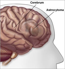
Low Grade Gliomas
Start with this review article (go here or here and see the NCCN site here). The grade and histology are critical in determining the treatment and prognosis for primary brain tumors. Low grade gliomas (grade 1 and 2) are more slow growing and are often treated with surgery.
The role of postOp radiation is somewhat controversial and is discussed below (see CancerNet, and the NCCN guidelines flow chart and the NCCN manuscript,) The treatment of high grade gliomas ( grade 3 anaplastic astrocytoma or grade 4 glioblastoma) are discussed elsewhere.
(Low grade gliomas have a different appearance on MRI or CT compared to glioblastoma see here, here, here, here and here).
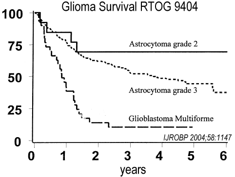
Survival tables are noted below. Old age has a negative impact on survival with low grade gliomas. In a large series from the Mayo clinic with patients over 55y the median survival was only 2.7 years (31%/5y and 19%/10y). Other Prognostic factors that affect survival are noted here, age and survival here, grade and survival go here, and the other studies below.
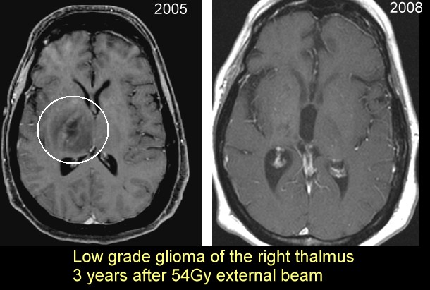
Studies using radiation (go here). Generally if radiation is used the dose is 54Gy in 30 factions as noted in the NCCN guidelines or the RTOG protocol below. The role of radiosurgery in this disease is controversial go here and here
Picture of a low grade glioma before after radiation go here. For more pictures of MRI images go here and here.
|
The target volume
is based on the T2 weighted images from the postoperative MRI scan (the preoperative
MRI scan can be used in patients who underwent biopsy only) and will include any T2
abnormality suspected of containing tumor plus a 2 cm margin (to block edge). If
the tumor has been completely resected, the target volume will be the surgical defect and
any T2 abnormality surrounding the surgical defect plus a 2 cm margin (to block edge). There will be no boost volume. The margin may be reduced to a 1 cm margin (to
block edge) around critical structures and/or natural barriers to tumor growth (e.g.
skull and tentorium). Treatment will be given in 1.8 Gy fractions (to isocenter),
1 fraction per day, 5 days per week and must begin within four weeks after randomization. The total dose will be 54 Gy in 30 fractions over approximately 6 weeks.
The target volume must receive 95-105% of the prescribed total dose to be per protocol (encompassed
by the 51.3 to 56.7 Gy isodose line). |
Survival | Astrocytoma | Astr-oligo | Oligodendroglioma |
median | 4.7y | 7.1y | 9.8y |
surv/5y | 46% | 63% | 73% |
surv/10y | 17% | 33% | 49% |
Author | Age | Median Survival |
Eyre | < 30y | not reached |
30 - 49y | 5.5 years | |
50 +y | 1.6 years | |
Medberry | <40 | 6.75 years |
40+y | 1.0 years | |
Piepmeier | < 40y | 8.7 years |
40 + y | 4.9 years | |
Shaw | < 35.5y | 6.3 years |
35.5y + | 4.2 years |
Survival (age less than) | Survival (age older than) |
6.75y (< 40y) | 1.0y (> 40y) |
8.7y (< 40y) | 4.9y (>40y) |
6.3y (< 35y) | 4.2y (> 35y) |
5.5y (< 35y) | 1.6y (> 35y) |
gross total resection | subtotal resection | biopsy only |
88% | 57% | |
85% | 64% | 43% |
80% | 50% | 45% |
Prognostic Groups as noted and below: Group I (KPS <70, age >40) MS 12 months; Group II (KPS > or =70, age >40, enhancement present) MS 46 months; Group III (KPS <70, age 18-40 or KPS > or =70 age >40, no enhancement) MS 87 months; Group IV (KPS > or =70, age 18-40) |