|
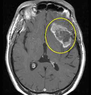
MRI Appearance of
Primary Brian Tumors (Gliomas, high grade glioblastoma and low grade gliomas.) GBM's are notoriously fast growing tumors as note here.
|
|
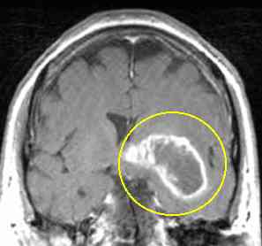
MRI Appearance of
Primary Brian Tumors (Gliomas, high grade glioblastoma and low grade gliomas.) GBM's are notoriously fast growing tumors as note here.
|
 |
 |
 |
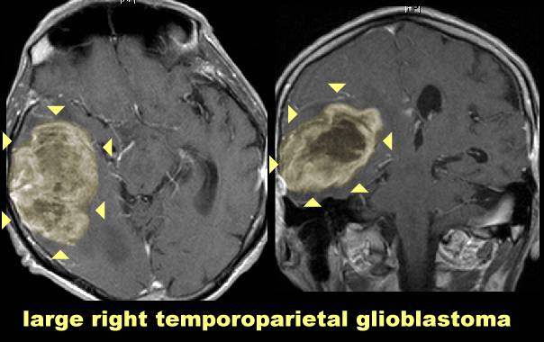
A grade 3 glioma (sometimes called anaplastic
astrocytoma) will have less necrosis in the center (compared to a grade 4 or glioblastoma)
but still look more abnormal (more enhancement) than a low grade (grade 1 or grade 2) . Low grade gliomas often recur, and at the time of the recurrence are higher grade (see pic of grade 1 that at the time of recurrence was grade 3) another low grade oligodendroglioma with anaplastic features on MRI here and course calcifications on CT here |
|
 |
note that a glioblastoma (above) looks more necrotic (gray area in the center, which contains dead or necrotic cells) than a low grade glioma (shown below:)
 |
Pictured at left is a low grade glioma ( Pilocytic Cerebellar Astrocytoma) with noncontrast CT on the left and axial T2- weighted MRI on the right.) from Harvard radiology (for more pics go here.) Picture of another low grade glioma |
![]()
 |
Malignant Glioma can appear as a complex cystic structure as noted on the left |
 |
occasionally a glioblastoma may appear as multiple
separate tumors and look metastatic disease (called a multicentric glioblastoma) |
