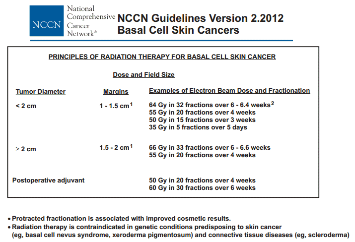
| Field Size: generally a margin of 1.0 - 1.5 cm is included around small tumors (up to 2cm) and 1.5 - 2.0 cm for larger lesions. For recurrent cancers or morphea basal cells an additional 0.5cm is added, and since electron beam field constrict, an additional 0.5cm. So the area treated may appear quite large to the patient (see dose and margins from NCCN). More dose information is here. In Europe faster regimens are used but often with inferior cosmetic results (go here). |
| Lesion | Energy | Dose | Fractions | Total Dose |
| 0.5 - 2.0 cm | 6- 9 MeV | 330 | 15-17 | 4950 - 5610cGy |
| >2 cm | 9 - 16 MeV | 275 | 20-22 | 5500-6050 |
| >2cm into bone | 9-20MeV | 200 | 34 -36 | 6800 - 7200 |
| Dose | Fractions | Days |
| Small area < 5cm2 | ||
| 20Gy | 1-2 | 1-2 |
| 30Gy | 5-10 | 5-14 |
| 40Gy | 10-16 | 16-28 |
| Larger area | ||
| 45Gy | 15-18 | 21-30 |
| 50Gy | 20-25 | 28-35 |
| 60Gy | 20-30 | 28-40 |
| We have generally found that older patients do not tolerate dose rates as fast as those described above an do better with three dose rates based on the size of the lesion and condition of the skin: |
| small lesions | 300cGy X 17 (5100cGy) |
| medium | 250cGy X 22 (5500cGy) |
| large or very delicate skin | 200cGy X 30 (6000cGy) |
What is the microscopic tumor extent beyond clinically delineated gross tumor boundary in nonmelanoma skin cancers?, 21 Choo . IJROBP 2005;621096-1099 A total of 71 lesions in 64 consecutive patients, selected for surgical excision with frozen-section-assisted assessment of resection margins, were accrued. The distance of microscopic tumor extension beyond a gross lesion varied from 1 mm to 15 mm, with a mean of 5.2 mm. A margin of 10 mm was required to provide a 95% chance of obtaining clear resection margins. The microscopic tumor extent was positively correlated with the size of gross lesion, but not with other variables. When RT is chosen as the therapeutic modality for nonmelanoma skin cancers, the determination of the area to be treated usually relies on visual delineation of the gross tumor and the inclusion of a margin of normal-appearing skin to account for microscopic tumor extent. The tissue volume that contains the subclinical microscopic tumor extent in addition to the gross tumor is equivalent to clinical target volume in the radiotherapy setting. Inadequate margin is associated with an increased risk of treatment failure, while excessive margin poses increased radiation morbidity. Therefore, a guideline for the margin of normal skin to be included within the RT volume would be very useful in clinical practice. In the RT setting, little information exists with regard to how much margin should be given beyond a gross tumor volume for the treatment of BCC or SCC of the skin. A limited number of surgical series evaluating the extent of excision required to obtain clear resection margin have provided some insight into this matter. Determination of RT treatment volume remains very much subjective, however, and is usually based on a physician’s clinical judgment. That is, to some extent, due to the nature of radiation oncology practice, in which a significant proportion of cases referred for consideration of RT are often more complex, such as a large, ill-defined, lesion or recurrence after initial surgical excision. Wolf reported in a prospective study on 117 cases of previously untreated, well-demarcated BCC that a minimum margin of 4 mm was necessary to totally eradicate the tumor in more than 95% of cases. In this study, Mohs micrographic surgery was used for assessment of microscopic extent of tumor beyond gross tumor. It is important to note that this study targeted only those lesions with clinically well delineated tumor borders and no previous treatment. In a similarly designed prospective study for SCC, Brodland reported that 4-mm margins were adequate for most SCC, while a minimum of 6-mm margins were recommended for high-risk tumors (such as a size >2 cm, high histologic grade, and invasion of subcutaneous tissue). In our series, nonmelanoma skin cancers have, on average, microscopic extension of 5.2 mm beyond the clinically visible tumor extent. However, extension could be as much as 15 mm. This magnitude was greater than those of the two surgical series described above. The primary reason for this variation lies most likely in the difference of case selection between the series. Our study, unlike the others, specifically targeted those tumors with poorly defined clinical borders, diameters >2 cm, or histopathologic features showing morpheaform or sclerotic patterns, and recurrence after previous treatment. In our series, 43.7%, 53.5%, and 52.1% were recurrent cancer, sclerosing type, and tumor >2 cm, respectively. These clinical and histologic features imply a higher likelihood of ill-defined tumor borders and thus an increased risk of underestimation of tumor extent. This speculation was further supported in that 70.4% of the lesions required more than one excision to obtain clear resection margins in our cohort. Another source of variation stems from potential imprecision in the way the distance of microscopic tumor extent beyond gross tumor was measured in our study. For example, if the plastic surgeon removed normal surrounding tissue more generously than would have been required, the margin of normal tissue required to obtain clear resection margins would be overestimated in our study. Our study is
consistent with the observation of other studies in that
larger lesions have greater subclinical
tumor extension. This observation must be taken into account in
treatment planning. Other variables such as histology, the number of
surgical attempts required to obtain clear margins, and the presence or
absence of previous treatment did not show statistically significant
correlation with the microscopic tumor extent in our cohort, although
there was a trend toward positive correlation. This finding may be in part
due to a small sample size of our study, resulting in insufficient power
to detect such correla Surgical margins for basal cell carcinoma.
Wolf. Arch Dermatol 1987; 123:340-4. Surgical margins for excision of primary cutaneous squamous cell carcinoma. Brodland. J Am Acad Dermatol 1992; 27:241-8.
Do plastic surgeons resect
basal cell carcinomas too widely? A prospective study comparing surgical
and histological margins
Br J Plast Surg.
2002;55:293–297 |