Home

Cancer

Cancer Types

Links

Site Map

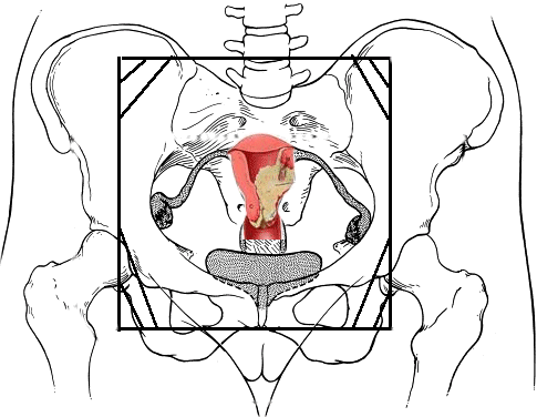
IMRT field (blue) surrounding the cervix cancer (red) and the involved lymph nodes (green)

GOG_logo.gif (7844 bytes)


We participate with the GOG (Gynecologic Oncology Group) which conducts national (international) research trials concerning the treatment of gynecologic cancer. For more information go here, and for typical radiation fields  used in the GOG protocols. More 
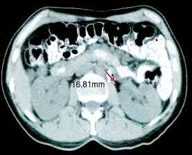
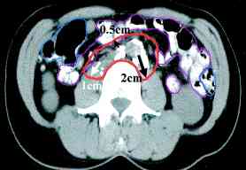
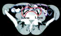
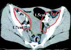
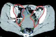
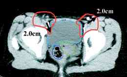
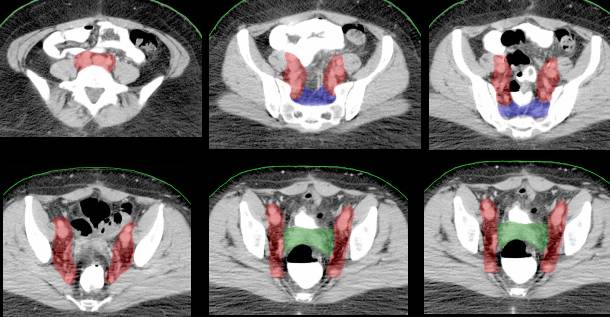
cervix ports, the node positions on CT scans (IJROBP 2002;54:1147)  (A) Furthest distance from lymph node to vessel wall. (B) Para-aortic lymph node CTV. (C) Common iliac lymph node CTV. (D) External iliac CTV, including lateral group. (E) External iliac CTV, including medial (obturator) group. (F) Inguinal lymph node CTV. CTV depicted by thick orange line. Small bowel demarcated by thin magenta, large bowel by thin blue, rectum by thin dark purple, bladder by thin turquoise, and uterus by thin yellow-green line. RTOG atlas here and here

AP 
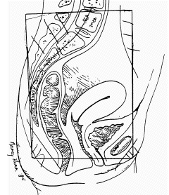
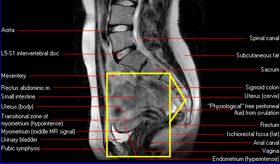
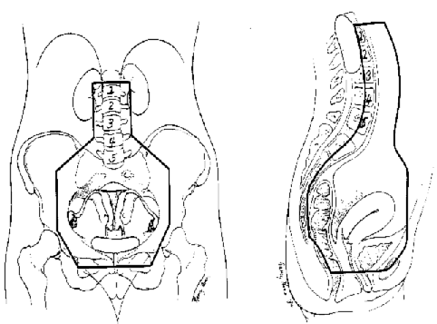
Pelvis Field (MRI appearance)
Lateral Pelvic Field (MRI appearance)
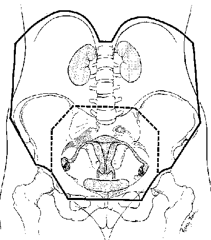
Para-Aortic Node Field
Inguinal/Vulva Field
Whole Abdominal Field


Note that internal radiation is commonly used as well as external beam. Internal radiation can be given with intracavitary Cesium sources (called LDR or low dose rate, go here) or HDR (high dose rate Iridium, go here.)  

Typical pelvic radiations from RTOG (0417 for advanced cervix cancer):

AP-PA Portals: Superior Border: A transverse line between L4 and L5 (or 1 cm margin on the superior extent of the uterus, whichever is higher). Inferior Border: Transverse line below the lowest extent of the obturator foramen or 3 cm below the most distal vaginal disease (whichever is most inferior), to include the introitus if necessary. Lateral Border: 2 cm lateral to widest true bony pelvic diameter. Custom Blocking: To shield small bowel and femoral heads while maintaining a margin of at least 1 cm from common iliac nodes and should not shield the obturator foramina (there should be at least 1.5 cm from the obturator foramen to block edge on the AP and PA fields).

Lateral Portals: Superior/Inferior Borders: Identical to AP-PA fields. Anterior Border: A straight line drawn 5 mm anterior to the symphysis pubis and at least 1 cm anterior to common iliac nodes at L4-L5. Posterior Border: In most cases, the entire sacrum should be included in the lateral fields. Posterior block should be designed so that the gross tumor is encompassed by at least 3 cm margins. In cases with small volume of disease, a line through the posterior sacrum may be used to include the cervical disease with a margin of 3-4 cm.

Reduced Fields: Parametrial/Nodal Boost (for Stage IIB and IIIB or involved pelvic lymph nodes. Note: Pelvic lymph nodes are considered those below the superior border of the sacrum. Those above the top of the sacrum are periaortic lymph nodes.