Radiation Dose Recommendations for Gamma Knife Radiosurgery (Pitt) (as of 2002)
Disorder Marginal Dose (50% IDL) Comments
AVM 15-25Gy varies by size and location
goal should be 24Gy (15-16 min) see AVM chart

Acoustic Neuroma 11-15Gy 13Gy  is usual (11-12 if 3cm)
12-13Gy at Cleveland

Meningioma 11-18Gy atypical (50-56Gy EBT + 12-16Gy)
malignant (60Gy EBT + 12-16Gy)
14-15Gy goal

Metastases 14-24Gy varies by size or combined with whole brain (see below)

Glial Tumors 12-20Gy EBT 55-60 Gy + 15.5 - 16Gy

Trigeminal Neuralgia 40Gy 80Gy maximum

Thalamotomy 60-70Gy 120-140Gy maximum

Pituitary 11-25Gy functional 16-25Gy
nonfunctional 12-16Gy
limit optic nerve and chiasm to 8Gy

The dose ranges above are from U Pitt. Other sites (e.g. Cleveland Clinic) have standard doses: acoustic neuroma of 12-13Gy, meningioma 14-15Gy if < 35mm and at least 5mm from the optic chiasm, and pituitary15-20Gy (higher if functional.) Volume is critical so in an AVM consider dose adjustments like: < 2cc (24Gy) 2.1- 5cc (22-23Gy) 5-8cc (21Gy) 8 - 13.5cc (18Gy) and > 13.5cc. The AVM (Pitt) data used 12Gy volume to predict toxicity and volumes up to 20cc had a low risk (< 10%) except brain stem sites.
See the tables on doses for single fraction or three fractions or five fractions

Brain Metastases Doses
Size Pit RTOG Other
2cm 20Gy 24Gy 24Gy
2.1-3cm 15-16Gy 18Gy 22-24
3.1-4cm 13.5 - 14Gy 15Gy 16Gy
whole + RS 16Gy   18Gy-20Gy

Karolinska Policy on Brain Mets
(assumes no whole brain/adjusts for critical structures)
Tumor Diameter Prescription Dose (40-60%IDL)
< 15mm 25Gy
15- 20mm 20-25Gy
20-25mm 20Gy
25-30mm 18Gy
>30mm don't treat


Some protocols for patients with brain metastases combine whole brain irradiation (WB) with radiosurgery as noted below:

RTOG 95-08 (for patient with 1-3 brain mets up to 4cm in size)  randomized patients between whole brain radiation alone (250cGy X 15 or 3750cGy) or WB followed by  radiosurgery the dose as noted:

2cm = 24Gy
2.1 - 3cm = 18Gy
3.1 - 4cm = 15Gy

ACOSOG Z0300, for patients with 1-3 brain mets (all less than 3cm)  all patients received radiosurgery and were then randomized to half of them receiving whole brain irradiation (250cGy X 12 or 3000cGy) which was given after the radiosurgery. The radiosurgery dose was adjusted lower for patients getting WB as noted below:
Radiosurgery Dose from ACOSOG Z0300
Tumor Size No Whole Brain Combined with Whole Brain
< 2cm 24Gy 22Gy
2 - 2.9cm 20Gy 18Gy


the dose for a brain metastases may be better chosen based on the volume rather than the diameter. For small volume lesions (up to 4-5 cc) full doses can probably be given (e.g. 22-24Gy, if over the motor strip 22Gy) for somewhat larger (5-8cc) the dose can be dialed down a bit (20-22Gy, but it may be useless to treat radioresistant mets to < 20Gy) for larger lesions (>8cc) then lower doses (16Gy) may be necessary. There are some studies with brain mets that showed better results with a dose of at least 20Gy in terms of  increased survival. There may be  still be a palliative benefit gained even if lower doses have to be used.
 
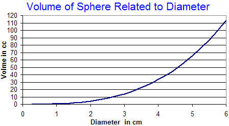
volume_sphere.gif (5445 bytes)
Diameter (cm) Volume (cc) (4/3 pi r^3)
0.25 .0082
0.5 .06545
.75 .2209
1.0 .5236
1.25 1.02265
1.5 1.767
1.75 2.806
2.0 4.1888
2.25 5.9641
2.5 8.18
2.75 10.889
3.0 14.137
3.25 17.974

Other dose guidelines from the Karolinska (as 2002)
  • GBM: 30Gy/10fx (EBT) + 15-18Gy (50% IDL) but 20% may require reoperation for radionecrosis, RTOG class III/IV may benefit
  • Low grade glioma: 10-12Gy (50% IDL) after surgery if residual
  • Meningioma:12-15Gy (50% IDL) lower dose if near critical structure, do not include dural tail
  • Malignant meningioma: 18Gy minimum , same dose/volume as mets
  • Acoustic neuroma: 12Gy (50% IDL) outline cochlea and plug to reduce fall-off, maximum volume 10cc
  • Pituitary: 12-15Gy (50% IDL) functional need higher dose, limit optic chiasm to 8Gy
  • AVM: 15-24Gy periphery, location more important than volume, cover entire nidus, embolize first if too big, limit brain stem to 15-18Gy
  • Trigeminal neuralgia:85Gy Dmax (4mm) shot placed at periphery and 50% ISD adjacent to brain stem
  • Epilepsy: 20-24Gy (50% IDL) / 4mm shot, collateral sulcus, parahypocampal area, limit BS to 12Gy and OC to 8Gy
  • Dose Limits to Critical Structure: Optic: 8Gy (6Gy if combined with external); Brain Stem: 12-18Gy, and Trigeminal: < 19Gy
  • Quality Assurance: the goal of planning as per RTOG 95-08 would be to have tumor coverage of 90% (Vpd minimum 80%) and Conformality Index (CI = PIV/TV) of < 2 (up to 3.5)

Radiation Dose by Tumor Size that will have a 1% (Kjellberg data) or 3% (integrated logistic formula) risk of radiation necrosis
Diameter (mm) Volume (cc) Gy (1% K) Gy (3% ILF)
12.5 1.02 27.5 34
15.0 1.77 25.0 29
17.5 2.81 22.5 23
20.0 4.10 20.0 18
22.5 5.96 18.7 16.5
25.0 8.18 17.5 14.5
27.5 10.89 16.5 13.5
30.0 14.14 15.0 13