|
|
|
|
|
|

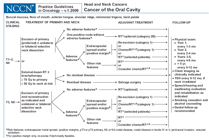
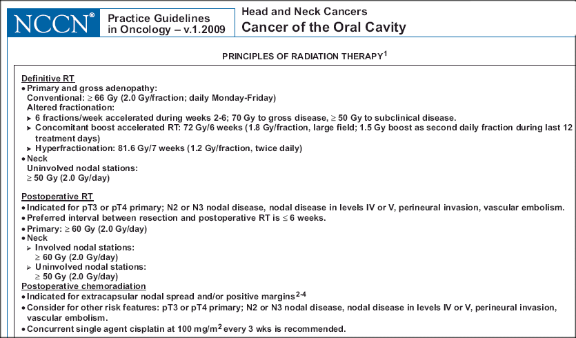
(also see NCCN guidelines for oral cavity cancers, early, advanced, most advanced and dose recommendations.)
Go here and here for some studies on outcome and treatment. Read review article here.
Squamous cell carcinomas arising in this area are usually well differentiated and are
frequently associated with areas of leukoplakia. Papillary, verrucous, and exophytic
mucosal growths are usually well differentiated with a low incidence of lymph node
metastases (i.e., 10% to 20% for T1 and T2 lesions).
Ulcerative, advanced tumors, which
are often associated with muscle invasion, have a higher propensity (60%) for lymph node
metastases.

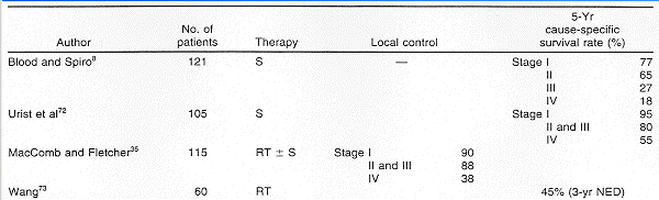
Buccal mucosa cancer � Buccal mucosa cancers are often neglected or misdiagnosed as an infection or consequence of trauma, and thus, rarely present as T1 lesions. Three-year disease-free-survival of 75 to 85 percent for stage I and 65 percent for stage II cancers have been reported
Surgery is typically preferred for buccal mucosa cancers, despite high local recurrence rates and technical challenges; postoperative radiation or chemoradiation is often necessary to optimize locoregional control. Exposure of the cancer can be difficult via a transoral approach, which makes it difficult to obtain clear radial margins in an en bloc fashion. Furthermore, the thin distance between the buccal mucosa and the buccal space permits early invasion to deep structures or to anterior cheek skin. When this happens, the surgeon must decide between taking a thin deep margin and risking recurrence or removing skin and reconstructing both inner and outer cheek surfaces. Although a more aggressive surgery, including exenteration of the buccal space and parotidectomy, may improve oncologic results, the disfigurement and morbidity of these procedures are considerable
Regardless of the depth of resection, the buccal mucosal surface must be aggressively reconstructed, and inadequate soft tissue coverage will result in severe, irreversible trismus. Therefore, many head and neck surgeons recommend free tissue transfer reconstruction (eg, radial forearm flap) for all but the smallest buccal cancers. Even with adequate reconstruction, aggressive rehabilitation must be instituted to optimize functional outcomes.
If RT is used, the initial treatment volume includes the primary tumor with at least 2 cm margins, and an intraoral stent may be used to displace the tongue and reduce the dose delivered to the contralateral side.