|
|
|
|
|
|
 |
||
|
|
|
|
|
|
|
|
|
||
|
|
HIGH-GRADE GLIOMA OVERVIEW
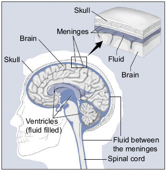
Primary brain tumors are cancers that originate in the brain. These tumors are very different from secondary brain tumors, which originally developed elsewhere in the body and spread (metastasized) to the brain. Primary brain tumors develop from glial cells. Glial cells provide the structural backbone of the brain and support the function of the neurons (nerve cells), which are responsible for thought, sensation, muscle control, and coordination. CLASSIFICATION OF PRIMARY BRAIN TUMORS Primary brain tumors are tumors that classified according to their appearance under the microscope. Gliomas are classified into four grades (I,II,III and IV), and the treatment and prognosis depend upon the tumor grade Grade I or II tumors are termed low-grade gliomas. The term malignant or high-grade glioma refers to tumors that are classified as:
HIGH-GRADE GLIOMA SYMPTOMS Gliomas cause symptoms by invading (growing) into and/or creating pressure in nearby normal brain tissue. The most common symptoms include:
Other common symptoms of brain tumors include memory loss, muscle weakness, visual symptoms, difficulty in using or understanding language, and personality changes. HIGH-GRADE GLIOMA TESTS Imaging studies — If your healthcare provider is concerned about your symptoms, s/he may recommend a scan of the brain. This can be done using MRI or CT. Both tests provide a very detailed image of the brain. However, a CT or MRI cannot determine for sure if a mass is a cancerous tumor. Biopsy — The only way to determine the type of tumor with certainty is for a neurosurgeon to remove a piece of the tumor (biopsy), usually during surgery. A pathologist will then examine the biopsy under a microscope. However, a biopsy may be done without surgery; this approach is preferred if the tumor is located within a critical area of the brain or if you are too sick for surgery. In these circumstances, a procedure called a stereotactic needle biopsy is used to take a sample of the tumor by inserting a needle through the skull into the brain itself. HIGH-GRADE GLIOMA INITIAL TREATMENT Treatment of a high-grade glioma includes measures to relieve symptoms and eliminate or reduce the tumor. This may include surgery, radiation, and/or chemotherapy. Everyone with high grade glioma is encouraged to participate in a clinical trial, if possible. Symptom management — Seizures and swelling in the brain (cerebral edema) can cause serious symptoms that may be life-threatening. Although treatment of the tumor may eventually alleviate these symptoms, treatments aimed at controlling the symptoms may be required:
To minimize side effects, the dose of dexamethasone (Decadron) is decreased gradually to the lowest level that controls symptoms.
Surgery — The initial treatment of high-grade glioma usually involves removing as much of the tumor as possible with surgery. The amount of tumor that can be removed is determined by the tumor's size and location, and by how much normal brain will be damaged as a result of surgery. The standard approach is to remove as much of the tumor as possible, while sparing areas of the normal brain that control critical functions such as speech or balance. Unfortunately, high-grade gliomas always have microscopic tumor cells that grow beyond the edge of the tumor. As a result, the tumor eventually regrows and few people with high-grade gliomas are cured with surgery alone. Radiation is typically recommended after surgery to kill any remaining tumor cells. Surgery may not be possible if the tumor is located in a part of the brain that controls critical functions or if you are in poor health. In these circumstances, radiation may be recommended as an alternative to surgery Radiation — Even when the entire tumor appears to have been removed, almost all high-grade gliomas eventually come back. This is because tumor cells have grown into the surrounding normal brain. Radiation therapy uses high energy x-rays to kill cancer cells and is usually recommended following surgery to kill remaining tumor cells. This treatment is called adjuvant radiation. Radiation can help to delay a recurrence of the tumor, allowing you to live longer. Radiation is generally given as a series of once daily treatments (called fractions) over several weeks. This approach helps to kill the greatest number of tumor cells and minimize side effects on normal brain cells. The area where the radiation is delivered (called the radiation field) is carefully calculated to include the smallest possible amount of normal brain as possible. Most brain tumors that grow back are within 2 cm (one inch) of the original tumor location. As a result, radiation is usually delivered to the "involved field" (the original area of the tumor plus a small margin) rather than the whole brain. Side effects — Radiation may kill normal brain cells as well as tumor cells, although tumor cells are somewhat more sensitive to the radiation. Damage to normal brain cells is often subtle, affecting mental sharpness and the ability to think and perform complex tasks (called cognitive impairment). Cognitive impairment tends to be more severe with larger radiation fields, tends to worsen over time, and is more of a problem in people who survive for several years after radiation treatments to the brain. It is not always possible to know if cognitive impairment is caused by radiation or a recurrence of the high-grade glioma. Chemotherapy — Chemotherapy refers to the use of medicines to stop or slow the growth of cancer cells. Chemotherapy works by interfering with the ability of rapidly growing cells (like cancer cells) to divide. Because most of an adult's normal cells are not actively growing, they are not affected by chemotherapy, with the exception of bone marrow (where blood cells are produced), the hair, and the lining of the gastrointestinal tract. Effects of chemotherapy on these and other normal tissues cause side effects during treatment. When used in combination with radiation therapy and surgery, chemotherapy may improve survival and quality of life in some patients with high-grade gliomas. The drug that are most widely used for high-grade glioma include temozolomide (Temodar®). Temozolomide (Temodar®) is usually taken by mouth for five consecutive days every four weeks. It is usually taken during and after radiation therapy. |