Cavernous Sinus Meningioma


Many tumors (particularly those involving the cavernous sinus) cannot be totally excised because of their relationship to vital neural or vascular structures. The rate of recurrence is markedly increased in these cases,: In a study of 581 patients undergoing initial resection for primary meningioma from 1978 to 1988, progression-free survival was higher in patients with gross total resection, compared to those with less than total resection at both five (88 versus 61 percent) and 10 years (75 versus 39 percent). In a second series of 935 patients who underwent surgery between 1953 and 1980, 15-year survival rate was 63 percent, a rate that was 78 percent of the normal life expectancy. Patients whose tumors were not completely removed had a 4.2-fold relative excess of death compared with those whose tumors were completely removed.

External beam RT decreased recurrence rates in patients with benign meningioma in whom gross total resection was not feasible. For such patients, subtotal resection combined with adjuvant radiation therapy can achieve results approaching those of total resection. RT can also be effective in meningiomas that are nonresectable. In a series of 132 patients, the 10-year local control rate in patients treated with total resection, subtotal resection plus RT, and subtotal resection alone were 77, 81, and 18 percent, respectively. Postoperative RT also improved local control when used with surgery to treat a first recurrence (10-year local rate 89 versus 30 percent with surgery alone). In a small 
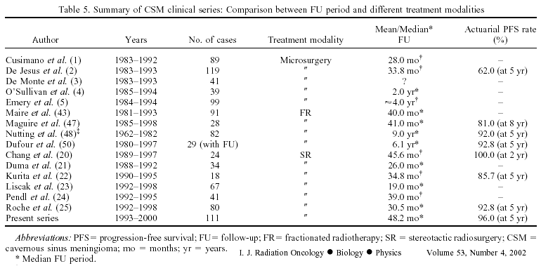
retrospective review of 31 patients with cavernous sinus meningiomas, 17 patients were treated with surgery and postoperative RT and 14 patients were treated with RT alone. Median dose was 52 Gy with standard fractionation and median follow up was 6.1 years. Ten-year progression free survival was 92.8 percent.

As note below, radiosurgery is particularly well suited for treating cavernous sinus meningioma, but
if the tumor is too close to the optic tract it may be better to use fractionated radiation (go here and here). For radiosurgery studies see below and here

gk_csm.jpg (13937 bytes)

The role of Gamma Knife radiosurgery in the management of cavernous sinus meningiomas


Antonio Nicolato
International Journal of Radiation Oncology.Biology.Physics, 2002; 53:4 : 992-1000


Axial MRI section showing a typical CSM before radiosurgery. (b) 13-month MRI FU after GK with significant shrinkage of the meningioma.

Study To evaluate the efficacy of Gamma Knife (GK) radiosurgery in terms of neurologic improvement and tumor growth control (TGC) in a large series of patients with cavernous sinus meningioma (CSM). The means and ranges of the parameters for the radiosurgical dose plan for the whole series were as follows: prescription isodose, 47.4% (30%–65%); prescription dose (PD), 14.8 Gy (11–22.5 Gy); maximal dose, 31.9 Gy (20.0–66.7 Gy); 
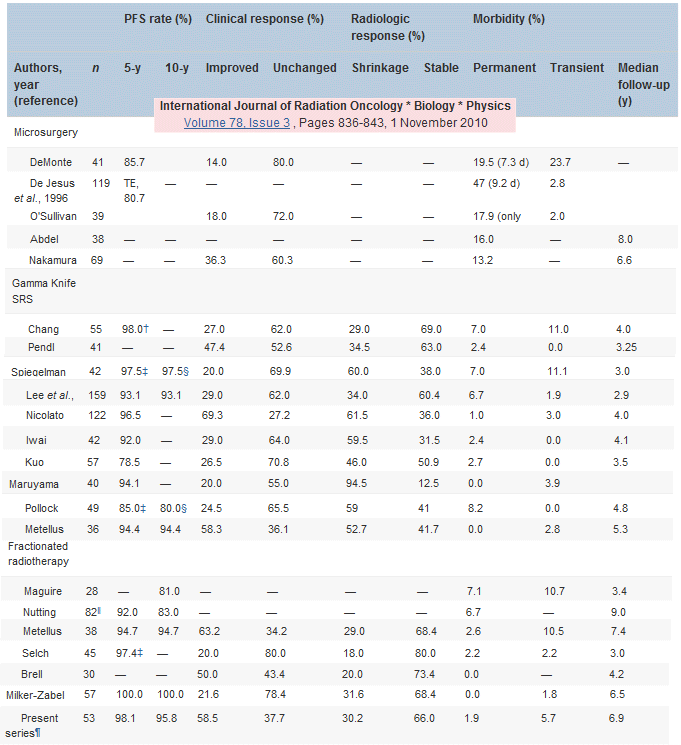
and number of isocenters, 9.3 (1–23).
     One hundred thirty-eight patients with CSM. Clinical conditions were improved or stable in 107/111 patients (96.5%). Neurologic recovery was observed in 76% of cases treated by GK alone and in 56.5% of adjuvant treatments. Adequate TGC was documented in 108/111 tumors (97%), with shrinkage/disappearance in 70/111 (63%) and no variation in volume in 38/111 (34%); the overall actuarial progression-free survival rate at 5 years was 96%.
Tumor size regression was observed in 79.5% of patients with FU >30 months, compared with 47.5% of patients with FU <30 months (p < 0.001). One hundred percent TGC was shown in treated patients with a CI [le] 1.5 (20/32), compared with 92% TGC in cases with a CI >1.5 (p < 0.15, NS). Radiosurgical sequelae were transient in 4/111 cases (3.5%) and permanent in one case (1%).

Conclusions : For the FU period of our series (median: >4 years), GK radiosurgery seems to be both safe (permanent morbidity 1%) and effective (96% neurologic improvement/stability, 97% overall TGC, 96% actuarial TGC at 5 years) and might be considered as a first-choice treatment for selected patients with CSM.

gk_nicolato.gif (23517 bytes)

1

Gamma knife radiosurgery in the management of cavernous sinus meningiomas.

Roche PH, Regis J, Dufour H, Fournier HD, Delsanti C, Pellet W, Grisoli F, Peragut JC.   J Neurosurg 2000 Dec;93 Suppl 3:68-73

Service de Neurochirurgie, CH St. Marguerite, Marseille, France.

Between July 1992 and October 1998, 92 patients harboring benign cavernous sinus meningiomas underwent GKS. The present study is concerned with the first 80 consecutive patients (63 women and 17 men). Gamma knife radiosurgery was performed as an alternative to surgical removal in 50 cases and as an adjuvant to microsurgery in 30 cases. The mean patient age was 49 years (range 6-71 years). The mean tumor volume was 5.8 cm3 (range 0.9-18.6 cm3). On magnetic resonance (MR) imaging the tumor was confined in 66 cases and extensive in 14 cases. The mean prescription dose was 28 Gy (range 12-50 Gy), delivered with an average of eight isocenters (range two-18). The median peripheral isodose was 50% (range 30-70%). Patients were evaluated at 6 months, and at 1, 2, 3, 5, and 7 years after GKS. The median follow-up period was 30.5 months (range 12-79 months). Tumor stabilization after GKS was noted in 51 patients, tumor shrinkage in 25 patients, and enlargement in four patients requiring surgical removal in two cases. The 5-year actuarial progression-free survival was 92.8%. No new oculomotor deficit was observed. Among the 54 patients with oculomotor nerve deficits, 15 improved, eight recovered, and one worsened. Among the 13 patients with trigeminal neuralgia, one worsened (contemporary of tumor growing), five remained unchanged, four improved, and three recovered.

Analysis of treatment outcome after stereotactic radiosurgery for cavernous sinus meningiomas.

Shin M, Kurita H, Sasaki T, Kawamoto S, Tago M, Kawahara N, Morita A, Ueki K, Kirino T.   J Neurosurg 2001 Sep;95(3):435-9

Department of Neurosurgery, The University of Tokyo Hospital, Japan.

The authors retrospectively reviewed their experience with 40 CS meningiomas treated with gamma knife radiosurgery. METHODS: Follow-up periods for the 40 patients ranged from 12 to 123 months (median 42 months), and the overall tumor control rates were 86.4% at 3 years and 82.3% at 10 years. When the tumor was completely covered with a dose to the margin that was higher than 14 Gy (Group A, 22 patients), no patient showed recurrence within the median follow-up period of 37 months. On the other hand, when a part of the tumor was treated with 10 to 12 Gy (Group B, 15 patients) or did not receive radiation therapy (Group C, three patients), the recurrence rates were 20% and 100%, respectively. Neurological deterioration was seen in nine patients, but all symptoms were transient or very mild. CONCLUSIONS: The data indicate that stereotactic radiosurgery can control tumor growth if the whole mass can be irradiated by dosages of more than 14 Gy. When optimal radiosurgical planning is not feasible because of a tumor's large size, irregular shape, or proximity to visual pathways, use of limited surgical resection before radiosurgery is the best option and should provide sufficient long-term tumor control with minimal complications.

Gamma knife radiosurgery of meningiomas in the cavernous sinus region.

Liscak R, Simonova G, Vymazal J, Janouskova L, Vladyka V.   Acta Neurochir (Wien) 1999;141(5):473-80

Department of Stereotactic and Radiation Neurosurgery, Hospital Na Homolce, Prague, Czech Republic.

For 6 years (1992-1998) we have treated 67 patients with cavernous sinus meningioma using the Leksell gamma knife in the Hospital Na Homolce, Prague. The age of the patients ranged between 19-82 years, median 57 years. Radiosurgery was the primary treatment in 64.2% of the patients, in the rest a microsurgical resection preceded. The volume of the tumour ranged from 0.9-31.4 cm3, median 7.8 cm3. The meningioma was distant from the optic tract in 58% of the cases, in 12% of the cases there was a contact with the tumour and the optic tract without its compression and in 30% of the cases there was a compression of the optic tract caused by the meningioma. The dose to the tumour margin ranged from 10-14 Gy, median 12 Gy. The follow up was available in 53 patients, in intervals of 2-60 months, median 19 months. There was no change in the tumour volume in 48% of the cases, in 52% of the cases a decrease of the tumour volume occurred. No increase of the tumour volume was observed. Clinical symptoms and signs improved in 35.8% of the patients, temporary morbidity was 3.8%. The mortality of the treatment was zero. Hitherto, the results of gamma knife radiosurgery of cavernous sinus meningioma have proved its safety and efficiency, although long term experience with a large group of patients is missing. Advances in neuroradiology and radiosurgical technique have allowed us to treat tumours with a closer contact to the optic tract and nerves compared with the past.

Fractionated stereotactic radiation therapy in the management of benign cavernous sinus meningiomas : long-term experience and review of the literature.

Milker-Zabel S,  Strahlenther Onkol. 2006 Nov;182(11):635-40

Department of Radiation Oncology, Radiotherapy, University of Heidelberg, 69120 Heidelberg, Germany. stefanie_milker-zabel@med.uni-heidelberg.de

PURPOSE: To analyze own long-term results with fractionated stereotactic radiotherapy (FSRT) in patients with benign meningiomas of the cavernous sinus and to review the literature on these rare lesions. PATIENTS AND METHODS: 57 patients were treated with FSRT for benign meningiomas of the cavernous sinus between 01/1990 and 12/2003 at the authors' institution. Histology was WHO grade I in 28/57 lesions, and undetermined in 29/57 lesions. 29 patients received radiotherapy as primary treatment, ten following surgery, and 18 patients were irradiated for recurrent disease. Median target volume was 35.2 cm3. Median total dose was 57.6 Gy with 1.8 Gy per fraction. 51/57 patients showed clinical symptoms before radiotherapy like reduced vision (n = 19), diplopia (n = 25), or trigeminal hyp-/dysesthesia (n = 17). RESULTS: Median follow-up period was 6.5 years. 50/57 patients were followed for > 36 months. Overall local tumor control was 100%. 39/57 patients had stable disease based on CT/MRI, while 18/57 had a partial remission of tumor volume. Overall survival for patients with WHO grade I meningiomas was 95.5% after 5 and 10 years. Two patients died 2.8 and 4.1 years after radiotherapy due to cardiac failure. In 11/57 patients, preexisting neurologic deficits improved. There was one patient with recurrent hyperlacrimation of one eye on the side of the irradiated meningioma. Three patients complained about subjective visual deterioration after FSRT without any objective findings in an ophthalmologic examination. No late toxicity RTOG >/= degrees III was seen. CONCLUSION: These data demonstrate that FSRT is an effective and safe treatment modality for local control of benign cavernous sinus meningiomas with a minimal risk of significant late toxicity.