| Stereotactic radiotherapy
for treatment of cavernous sinus meningiomas Selch MT, International Journal of Radiation Oncology*Biology*Physics 01 May 2004 (Vol. 59, Issue 1, Pages 101-111) |
||
|
||
| Despite significant advances in
neuroimaging and microsurgical techniques, meningiomas of the cavernous sinus continue to
represent a neurosurgical challenge. The preoperative assessment demonstrates total
encasement or narrowing of the intracavernous portion of the internal carotid artery in
63% of patients. Furthermore, although the microscopic appearance of meningioma is
typically benign, careful histopathologic analysis has demonstrated infiltration of
adjacent vascular structures and cranial nerves by cavernous sinus lesions. Consequently, the reported rates of gross total resection for cavernous sinus
meningiomas vary from 12% to 84%. Local recurrence is reported in 9.6–25% of patients
despite confirmed total resection of cavernous sinus meningioma on imaging. Local
progression is even more frequent after subtotal removal of meningioma. In a series of 119
cavernous sinus meningiomas, DeJesus reported a 5-year relapse-free survival rate of 81%
after complete resection compared with 62% after incomplete tumor removal. Attempted
removal of cavernous sinus meningiomas has been associated with serious morbidity. The rate of exacerbation or induction of cranial neuropathy resulting
from cavernous sinus surgery varies from 6% to 18%. In addition, hemorrhage and
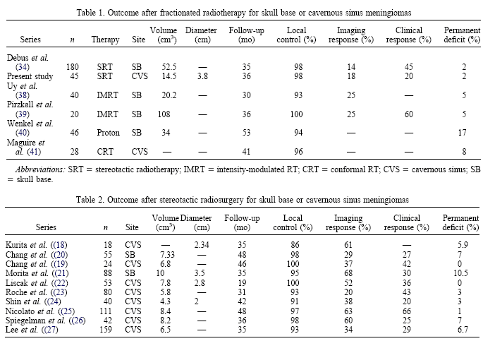
cerebrospinal fluid leak have been reported in 5% and 21%, respectively. Conventional external beam radiotherapy (RT) has been demonstrated to be an effective adjunctive therapy after subtotal resection of intracranial meningioma and a useful primary treatment modality for unresectable or inoperable tumors. Several authors have demonstrated relapse-free survival rates after external beam RT for cavernous sinus meningiomas that equal or exceed the rates achieved by radical resection). External beam RT, however, may occasionally produce long-term optic, pituitary, or cognitive dysfunction Stereotactic radiosurgery (SRS) is a method for delivering a large, single dose of radiation to an intracranial site. The inherently steep dose gradient produced by SRS affords physical protection of the normal tissue adjacent to the target lesion. SRS has proved effective for a variety of benign central nervous system disorders, including meningiomas. Local control rates of 86–100% have been reported after either gamma-knife or linear accelerator-based SRS for selected patients with cavernous sinus meningiomas Despite the dosimetric advantages of SRS, use of this technique for cavernous sinus meningiomas has resulted in a 1.5–10.5% serious morbidity rate, predominantly affecting the cranial nerves. Morbidity after SRS is strongly associated with the tumor volume and location). For these reasons, application of SRS to cavernous sinus meningiomas has generally been restricted to tumors <3 cm in greatest dimension and located several millimeters from the optic apparatus. Stereotactic RT (SRT) combines the physical dose localization advantages of SRS with the radiobiologic benefits of dose fractionation. Unlike SRS, the treatment planning fiducial system and patient immobilization device can be applied noninvasively. The accuracy of patient repositioning during SRT has been documented. A total fractionated dose of ionizing radiation, known from conventional RT experience to be effective for meningioma and tolerated by the central nervous system, can be delivered by the SRT technique. SRT has proved safe and efficacious for a variety of skull base tumors, including meningiomas. We report the results of SRT using a dedicated linear accelerator equipped with a micromultileaf collimator (MMLC) for 45 patients with meningiomas of the cavernous sinus. Most of the patients had tumors considered ineligible for treatment by SRS. To assess the safety and efficacy of
stereotactic radiotherapy (SRT) using a linear accelerator equipped with a micromultileaf
collimator for cavernous sinus meningiomas.
|