 |
Testicular cancer, although an
uncommon malignancy, is the most frequently occurring cancer in young men. In the year
2000, an estimated 7,400 cases of testicular cancer will be diagnosed. For unknown
reasons, the incidence of this cancer has increased since the turn of the century, from 2
cases per 100,000 population in the 1930s to 3.7 cases per 100,000 from 1969 to 1971 to
4.1 cases per 100,000 from 1983 to 1987.
Most testicular tumors are of germ-cell
origin. These cancers are uniquely sensitive to chemotherapy and are considered the model
for the treatment of solid tumors. Perhaps the most controversial area in the management
of germ-cell tumors is the proper approach to early-stage disease; ie, surveillance vs
primary lymphadenectomy (for nonseminoma germ-cell tumors) or radiation therapy (for
seminomas). In advanced disease, chemotherapy plays an essential role, but novel treatment
regimens are currently being evaluated through multi-institution clinical trials |
CT Scans are important in staging
these men to look for lymph nodes in the abdomen or pelvis or spread to the lung or
mediastinum. Tumor markers are also important:
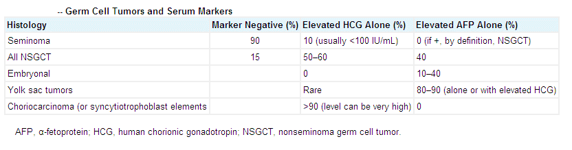
The serum
tumor markers
alpha-fetoprotein (AFP), lactate dehydrogenase (LDH), and human chorionic gonadotropin
(hCG) are of critical importance in diagnosis, prediction of prognosis, and assessment of
treatment outcome. They should be determined before, during, and after treatment as well
as throughout the followup period. Alpha-fetoprotein is a serum tumor marker produced by
nonseminomatous cells (embryonal carcinoma, yolk-sac tumor) and may be seen at any stage.
An approximate half-life of alphafetoprotein is 5-7 days. A nonseminoma, therefore, is
associated with elevated serum concentrations of AFP. An elevated serum concentration of
hCG (half-life is approximately 1-3 days) may also be present with both seminomatous and
nonseminomatous tumors.
Seminomas are occasionally associated
with an elevated serum concentration of hCG, but not an elevated concentration of AFP.
Nonseminoma is the more clinically aggressive tumor. When both a seminoma and nonseminoma
elements are present, management follows that for a nonseminoma.
Epidemiology
Age Testicular cancer can
occur at any age but is most common between the ages of 15 and 35 years. There is a
secondary peak in incidence after age 60. Seminoma is the most common histology in the
older population but is rare in those under age 10.
Race Testicular cancer is rare
in African-Americans, occurring at a rate of 0.9 per 100,000 population. Incidence of this
cancer has increased in whites during the 20th century but has remained flat in
African-Americans.
Geography Denmark has the
highest incidence of testicular cancer and the Far East, the lowest.
Primary site Germ-cell tumors
present most commonly in the testis (90%) and only infrequently in extragonadal sites
(10%). The most common extragonadal sites (in decreasing order of frequency) are the
retroperitoneum, mediastinum, and pineal gland. Many patients presumed to have a primary
retroperitoneal germ-cell tumor may have occult germ-cell tumors of the testicle. This
possibility should be evaluated with testicular ultrasound, especially when the
retroperitoneal tumor is predominantly one-sided.
Survival The 5-year survival
rate for all patients with a germ-cell tumor is ~ 95%. Cure rates are highest for
early-stage disease, which is treated primarily with surgery or radiation therapy (early
seminoma), and lower for advanced disease, for which chemotherapy is the primary therapy.
Etiology and risk factors
The specific cause of germ-cell
tumors is unknown, but various factors have been associated with an increased risk of this
malignancy.
Prior testicular cancer
Perhaps the strongest risk factor for germ-cell tumors is a previous history of testicular
cancer. Approximately 1%-2% of patients with testicular cancer will develop a second
primary in the contralateral testis over time. This represents a 500-fold increase in
incidence over the normal male population.
Cryptorchidism Patients with
cryptorchidism have a 20- to 40-fold increased risk of developing germ-cell tumors,
compared with their normal counterparts. Orchiopexy, even at an early age, appears to
reduce the incidence of germ-cell tumor only slightly (if at all). Of note, in ~ 10% of
patients with cryptorchidism who develop germ-cell tumors, the cancer is found in the
normally descended testis. Biopsies of nonenlarged cryptorchid testes demonstrate an
increased incidence of intratubal germ-cell neoplasmľa presumed precursor lesion.
Genetics Klinefelter’s
syndrome (47XXY) is associated with a higher incidence of germ-cell tumors, particularly
primary mediastinal germ-cell tumors. In addition, patients with Down’s syndrome have
been reported to be at increased risk for germ-cell tumors. Also thought to be at greater
risk are patients with testicular feminization, true hermaphrodites, individuals with
persistent müllerian syndrome, and persons with cutaneous ichthyosis.
Family history Although
familial testicular cancer has been observed, the incidence among first-degree relatives
remains low. One investigator, however, reported a 6-fold increased risk among male
offspring of a testicular cancer patient.
Environment Numerous
industrial occupations and drug exposures have been implicated in the development of
testicular cancer. Although exposure to diethylstilbestrol (DES) in utero is associated
with cryptorchidism, a direct association between DES and germ-cell neoplasm is weak at
best.
At least two reports have suggested
an increased risk of testicular cancer among individuals exposed to exogenous toxins, such
as Agent Orange and solvents used to clean jets.
Prior trauma, elevated scrotal
temperature (secondary to use of thermal underwear, jockey shorts, and electric blankets),
and recurrent activities, such as horseback riding and motorcycle riding, do not appear to
be related to the development of testicular cancer.
No supporting findings substantiate a
viral etiology.
Diagnosis
Ultrasonography can reliably
identify masses within the testis. In virtually all patients, ultrasonography can
distinguish a testicular from an extratesticular mass and may detect lesions that are not
palpable on physical examination. Ultrasound findings cannot consistently differentiate
benign from malignant tumors of the testis (95% of such masses are malignant). Most
patients with testicular cancer, especially seminoma, have hypoechoic lesions compared to
adjacent tissue. Nonseminomatous tumors, however, may have mixed signals, including
hyperechoic masses, which are commonly seen with teratoma.
Serum markers Serum levels of
b-subunit human chorionic gonadotropin (b-HCG) and a-fetoprotein (AFP) are elevated in ~
80%-85% of patients with extensive germ-cell tumors. Patients with pure seminoma may have
elevated levels of b-HCG but not AFP (elevated AFP indicates the presence of
nonseminomatous germ-cell elements). False-positive b-HCG levels can be seen in patients
who have hypogonadism (cross-reactivity with luteinizing hormone) or who use marijuana,
whereas AFP may be elevated in patients with liver dysfunction or hepatitis.
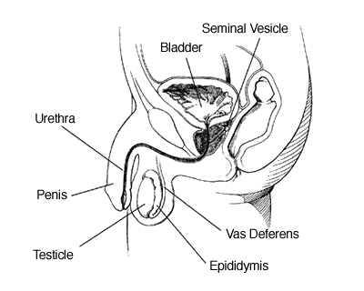
Inguinal orchiectomy When a
testicular mass is discovered, the patient should undergo an orchiectomy through an
inguinal incision.
Trans-scrotal incisions or
biopsies should not be performed, as they ultimately lead to aberrant lymphatic
drainage from the tumor.
Pathology
Germ-cell tumors are classified into
two broad histologic categories: seminoma and nonseminomatous germ-cell tumor. Patients
with seminoma who have increased AFP or any focus of nonseminomatous germ-cell tumor
components (including teratoma) are considered to have a nonseminomatous germ-cell tumor.
Seminoma
Seminoma is the most common single
histology, accounting for ~ 30% of all germ-cell tumors. Up to 10% of seminomas have focal
syncytiotrophoblast cells, thought to be the source of b-HCG in some cases.
Spermatocytic seminoma, a rare
subset of germ-cell tumors, often grows to large size, occurs almost exclusively in men
over 50 years old, and rarely metastasizes.
Nonseminoma
Embryonal carcinoma is composed of
large pleomorphic cells with different architectural patterns. This tumor may be
associated with an elevation of serum b-HCG and/or AFP.
Endodermal sinus tumor (yolk sac
carcinoma) is the most common testicular tumor seen in infants and young children.
Like embryonal carcinoma, yolk sac tumor has a variety of architectural patterns. This
tumor is associated with elevated serum AFP.
Choriocarcinoma, as a pure
entity, is one of the least common germ-cell tumors. These tumors have a great propensity
for hematogenous spread, often skipping the retroperitoneum. Choriocarcinoma is associated
with increased serum b-HCG.
Teratoma is a generally benign
tumor with elements from each of the germ layers (ectoderm, mesoderm, and endoderm).
Teratoma is uncommonly seen as the sole histology in primary tumors, but it is frequently
associated with other histologic elements, including those mentioned above. Of patients
with residual disease following chemotherapy for nonseminomatous germ-cell tumor, ~ 45%
have teratoma in resected specimens.
A subset of patients with immature
teratoma that contains non-germ-cell histologies (eg, sarcoma, adenocarcinoma) has been
reported. In contrast to most teratomas, these tumors may grow locally and can be lethal.
In addition, late recurrences of both teratoma and carcinoma have been reported in
patients with teratoma. Serum markers are normal in patients with pure teratoma.
Treatment
surgical treatment of stage I or
II disease
Initial intervention for testicular
cancer is radical inguinal orchiectomy. Orchiectomy may be deferred temporarily in
patients with advanced-stage disease in whom the diagnosis of nonseminomatous germ-cell
tumor can be made on clinical grounds (elevated markers). In such patients, an orchiectomy
must be performed later, as there is incomplete penetration of chemotherapy into the
testes.
Further therapy hinges on the
pathologic diagnosis. In general, pure seminomas (normal AFP with or without elevated
b-HCG) are treated with radiotherapy and/or chemotherapy, whereas most nonseminomas are
treated with surgery and/or chemotherapy.
Radiation therapy for Stage I or
II seminomas
Seminomas of the testes are
exquisitely sensitive to radiation. This characteristic, combined with their predictable
lymphatic spread, make these cancers amenable to radiotherapy. Since low radiation doses
are used, acute and late side effects are few.
Stage I disease
Prophylactic radiotherapy vs
chemotherapy vs surveillance Primary lymphatic drainage of the testis is to the
para-aortic lymph nodes from the level of the renal vessels to the bifurcation of the
aorta. While ipsilateral pelvic nodal failures and, to a much lesser extent, inguinal
failures have been reported following tumor resection by inguinal orchiectomy in patients
with stage I disease, these sites have a much lower risk of failure than do the
para-aortic lymph nodes. Based on surveillance data, the overall incidence of disease
failure without radiotherapy is 15%-27% (median, 20%), whereas only 2%-5% (median, 3%) of
patients who are treated with radiotherapy relapse.
Although surveillance would appear to
be a reasonable approach for patients with stage I disease, since 80% of these patients
will be treated unnecessarily, follow-up is problematic because progression usually is not
associated with symptoms until the tumor burden is large. Surveillance requires frequent
abdominopelvic CT scans and chest x-rays for 4-5 years. Despite excellent salvage rates
reported in patients who relapse while undergoing surveillance (initial salvage rates of
approximately 90%, with ultimate salvage rates after relapse of approximately 95%), most
groups have discontinued surveillance protocols in lieu of prophylactic radiotherapy to
the draining lymphatics or chemotherapy. Those patients who do develop a recurrence
usually receive 4 cycles of etoposide plus Platinol (EP).
Several phase II trials have
evaluated 1-2 cycles of carboplatin (Paraplatin) for prophylactic treatment of stage I
seminona. The preliminary results are comparable to radiotherapy in terms of recurrence
rates. Late toxicity data are lacking, and prophylactic carboplatin therapy is not
considered to be a standard approach in this country.
Radiation fields and doses The
radiotherapy portals typically include the para-aortic lymph nodes from T10-L5 and the
ipsilateral hemipelvis, including the inguinal scar. However, recent studies that reduced
the size of para-aortic fields and omitted hemipelvis radiation in selected patients (eg,
those with no prior orchiopexy or other pelvic, inguinal, or scrotal surgery) are
encouraging. Pelvic and/or inguinal failures occurred in < 5% of these patients. The
smaller treatment volume reduces the dose to the remaining testicle and probably the risk
of second malignancy.
The hemiscrotum is usually treated if
the tumor penetrated the tunica albuginea, a trans-scrotal incision was performed, or
orchiopexy was done for cryptorchidism. Although treatment of the hemiscrotum for these
reasons remains a standard practice, it has been questioned since the incidence of scrotal
failure is low even in the presence of these risk factors. In fact, some surgeons advocate
trans-scrotal exploration to rule out benign lesions.
The sites listed above are treated
with 25-30 Gy over 3-3.5 weeks, although some data suggest that 20 Gy is sufficient. The
5-year actuarial rate of disease freedom using such techniques is 97% and the rate of
overall survival is nearly 100% since the availability of platinum-based chemotherapy for
salvage. The few failures observed following radiotherapy most often occur in the next
echelon of lymph node drainage sites, such as the mediastinum or left supraclavicular
fossa.
Side effects The acute side
effects of radiotherapy are limited to nausea, vomiting, and diarrhea, all of which
usually can be readily controlled with medication. Late toxicities are rare, although
peptic ulcers (~ 5%) and marginally higher rates of second malignancies have been
reported.
Permanent infertility from scattered
radiation to the contralateral testis is uncommon, whereas prolonged aspermia for over 1
year may occur, especially with irradiation of the hemiscrotum. Nevertheless, sperm
banking is recommended for those concerned about childbearing.
Stage II disease
The majority of patients with
infradiaphragmatic para-aortic and/or pelvic adenopathy < 5 cm are treated with
radiotherapy alone, while those with larger lymph node metastases are treated with
platinum-based chemotherapy. Some advocate a 10-cm adenopathy cut-off point for deciding
whether to use radiotherapy or chemotherapy. Among patients who are candidates for
radiotherapy, it is essential that renal function be preserved in case chemotherapy is
necessary for salvage treatment.
Radiation fields The
radiotherapy fields are similar to those used for stage I disease except that the fields
are widened to include any para-aortic or pelvic adenopathy with a 2- to 3-cm margin. In
the past, mediastinal and supraclavicular treatment was standard in patients with stage II
disease. However, data from several series revealed only a 3% rate of
mediastinal/supraclavicular relapse. In addition, late cardiac toxicity has been reported.
Although treatment to these sites has largely been abandoned in these cases, one report
indicates that the rate of failures in the left supraclavicular fossa is greater than was
previously believed.
The actuarial rate of freedom from
disease at 5 years for patients with para-aortic adenopathy < 5 cm is ~ 90%, vs 85% for
those with adenopathy > 5 cm and < 10 cm. Most of the data on the latter group of
patients are from older studies in which patients often received prophylactic mediastinal
and supraclavicular irradiation, and outcome may be poorer without such treatment.
However, this approach has been abandoned today in view of the excellent salvage rates
with chemotherapy.
Radiation doses The involved
areas are treated with 30-35 Gy and the uninvolved areas, with 25-30 Gy. There is no
evidence of a dose-response effect above 25 Gy for uninvolved areas and above 30 Gy for
involved areas. Failures within the irradiated volume are anecdotal.
Medical Treatment of Stage II
nonseminomas
Over the past several years, the
threshold for primary surgery in patients with stage II disease on CT scans has changed.
At present, masses > 3 cm in greatest cross-sectional diameter are generally handled
primarily with chemotherapy. For patients with tumor sizes Ł 3 cm, primary RLND is
considered the standard approach. It should be noted that up to 25% of patients with
enlarged lymph nodes on CT scans will be found to have pathologic stage I
(false-positives) disease by RLND.
Adjuvant chemotherapy for
nonseminomas
The risk of systemic recurrence is
5%-10% in patients with pathologic stage I nonseminoma, 15%-30% in those with completely
resected stage IIa (N2a) disease, and 30%-50% in those with stage IIb (N2b) disease.
Recurrence usually occurs in the lungs within the first 24 months after surgery. The risk
of retroperitoneal recurrence in patients with stage I, IIa, or IIb disease is < 1%
after a properly performed RLND.
Following RLND, patients with
complete resection of stage II disease can be considered candidates for adjuvant
chemotherapy.
The decision of whether or not to
prescribe adjuvant therapy following lymph node dissection is somewhat arbitrary, and
often depends on the patient’s social circumstances and likelihood of adhering to
close follow-up. A patient with completely resected carcinoma who undergoes an RLND has a
70% chance for cure; thus, the majority of patients will never need chemotherapy. However,
these patients must be monitored carefully with chest x-rays and serum marker
determinations every month for 1 year, every 2 months for an additional year, and then
every 6 months for the next 3 years. (CT scanning is not performed routinely unless
clinically indicated.) The 30% of patients followed in such a manner who do develop a
recurrence will present with a tumor of low volume (eg, small pulmonary metastases or
elevated serum markers); nearly 100% of these patients should be cured with appropriate
systemic therapy.
However, some patients with resected
stage II disease elect to receive adjuvant chemotherapy to minimize the risk of cancer
recurrence. For such therapy, 2 cycles of BEP (bleomycin, 30 IU/wk ×8; etoposide, 100
mg/m˛ on days 1-5 and 29-34; and Platinol, 20 mg/m˛on days 1-5 and 29-34) are
recommended.
It should be emphasized that in a
patient who agrees to close follow-up, the chance of dying of cancer should be negligible
in either scenario. For patients who have persistently elevated or increasing serum
markers following RLND or who have undergone an incomplete lymph node dissection, 3 cycles
of BEP are indicated.
TREATMENT OF Stage III Disease
Seminomas
Chemotherapy is the treatment of
choice for patients with stage III seminomas. The management of patients with bulky
disease after chemotherapy (residual mass > 3 cm) is somewhat controversial.
Investigators at Memorial Sloan-Kettering suggest that such patients require consolidation
with radiotherapy or surgical removal of radiographically evident disease. More recent
data from the Royal Marsden Hospital report a relapse rate of 10%-15% in patients with
residual masses with or without post-chemotherapy surgery or radiotherapy, supporting the
practice of observation in patients with residual disease following chemotherapy.
Nonseminomas
As mentioned above, patients with
nonseminomas being treated with chemotherapy can be classified as having good- or
poor-risk disease.
Good-risk nonseminomas In
patients with good-risk nonseminomas, 3 cycles of BEP (bleomycin, 30 IU/wk, and etoposide,
100 mg/m˛, both on days 1-5, plus Platinol, 20 mg/m˛ on days 1-5) given every 3 weeks
or, alternatively, 4 cycles of etoposide plus Platinol without bleomycin (EP; at the same
dosages) appear to yield equivalent results. Over 90% of good-risk patients should be
cured with these therapies.
Post-chemotherapy resection If a
patient has persistent radiographic disease with normal serum markers 4-6 weeks following
chemotherapy for a nonseminomatous germ-cell tumor, surgical resection should be performed
when possible.
Post-resection chemotherapy Histologic
examination of residual disease will reveal necrotic fibrous tissue in ~ 45% of such
cases, benign teratoma in ~ 45%, and persistent carcinoma in ~ 10%-15%. If persistent
carcinoma is detected in the resected specimen, 2 additional cycles of cisplatin plus
etoposide should be administered. For patients with complete resection of mature and
immature teratoma or necrosis, no additional therapy is needed.
Poor-risk nonseminomas A
cohort of patients with disseminated germ-cell tumors present with very advanced or
poor-risk disease. “Poor risk” has been variously defined but represents a
patient population with a cure rate of Ł 50% with standard cisplatin-based combination
chemotherapy.
Prior to the use of cisplatin-based
chemotherapy, radiation had been used in the treatment of minimal residual nonseminoma
following surgical dissection. The doses required for local control were 40-50 Gy. Such
treatment has been supplanted by chemotherapy. Radiation is useful in the treatment of
metastatic nonseminoma to the brain.
Chemotherapy During the last
several years, several trials have evaluated a variety of combination regimens in patients
with poor-risk disease. These include the use of high-dose cisplatin (40 mg/m˛ on days
1-5) or VIP (VePesid, ifosfamide, and Platinol). However, to date, none of these regimens
has demonstrated superiority over 4 cycles of BEP.
An ongoing intergroup trial is
currently comparing the role of high-dose chemotherapy with autologous bone marrow
transplantation (BMT) as part of induction chemotherapy vs standard BEP in patients with
poor-risk disease.
Post-chemotherapy resection The
ultimate goal of combination chemotherapy in these patients is resolution of all
radiographically visible disease and normalization of tumor markers. If residual
radiographic abnormalities persist in the lungs and/or abdomen, surgical resection of
residual disease is indicated.
A post-chemotherapy retroperitoneal
lymph node resection must clear the region of residual disease. In general,
post-chemotherapy resections are extremely difficult and incomplete resections are
unacceptable. After the retroperitoneum is cleared of persistent radiographic disease,
persistent pulmonary lesions are resected. In cases with residual disease in the
retroperitoneum and thorax, the RPLND should be performed first. If necrosis is found, the
disease within the chest can be observed. If teratoma or cancer is noted, the
supradiaphragmatic disease should be resected.
Complicating factors associated with
post-chemotherapy resections include the risk of oxygen toxicity secondary to bleomycin,
as well as intense fibrosis and adherence of residual disease to the aorta and other vital
retroperitoneal organs. Inspired oxygen levels must remain below 35% to prevent
bleomycin-related acute respiratory distress syndrome, which has a fatality rate ł 50%.
After successful resection, the only
visible structures remaining should include the back muscles, nerves, anterior spinous
ligament, aorta, IVC, renal vessels, kidneys, and ureters. Up to 20% of patients with
advanced abdominal disease may require resection of a kidney or even the IVC. Operative
mortality in centers with experience performing resections of these advanced-stage tumors
should be < 2%.
Post-resection chemotherapy As
mentioned, 2 additional cycles of chemotherapy are indicated for patients with persistent
viable carcinoma in the resected specimen. |