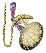

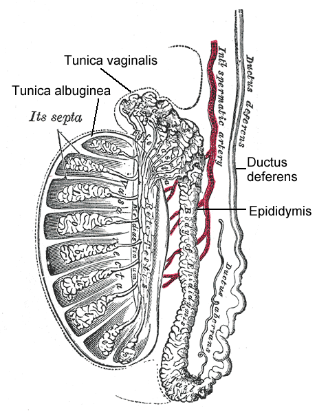
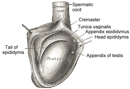
The testes
are 4 to 5 cm long, 3 cm wide, and 2.5 cm deep
and have a volume of 30 mL. They are enclosed
in a tough capsule comprising (1) the visceral
tunica vaginalis; (2) tunica albuginea, with
collagenous and smooth muscle elements; and (3)
the tunica vasculosa. The epididymis
attaches to the posterolateral aspect of the
testis. Beneath it, the tunica albuginea
projects inward to form the mediastinum testis,
the point at which vessels and ducts traverse
the testicular capsule. Septa radiate from the
mediastinum to attach to the inner surface of
the tunica albuginea to form 200 to 300
cone-shaped lobules, each of which contains one
or more convoluted seminiferous tubules. Each
tubule is U-shaped and has a stretched length of
nearly 1 m. Interstitial (Leydig) cells lie in
the loose tissue surrounding the tubules and are
responsible for testosterone production. Toward
the apices of the lobules, the seminiferous
tubules become straight (tubuli recti) and enter
the mediastinum testis to form an anastomosing
network of tubules lined by flattened
epithelium. This network, known as the rete
testis, forms 12 to 20 efferent ductules and
passes into the largest portion of the
epididymis, the caput. Here, the efferent
ductules enlarge, become more convoluted, and
form conical lobules. The duct from each lobule
drains into a single epididymal duct, which
winds approximately 6 m within the fibrous
sheath of the epididymis to form its body and
tail. As the duct approaches the tail, it
thickens and straightens to become the vas
deferens.
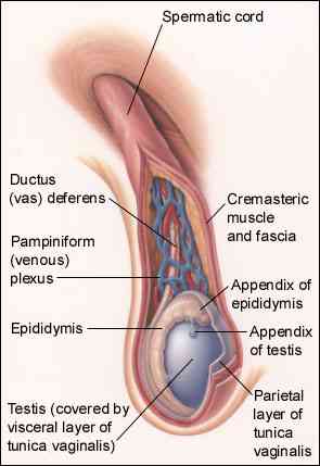
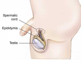
The spermatic cord is composed of the vas deferens, testicular vessels, and spermatic fasciae
