SKULL-BASE METASTASES
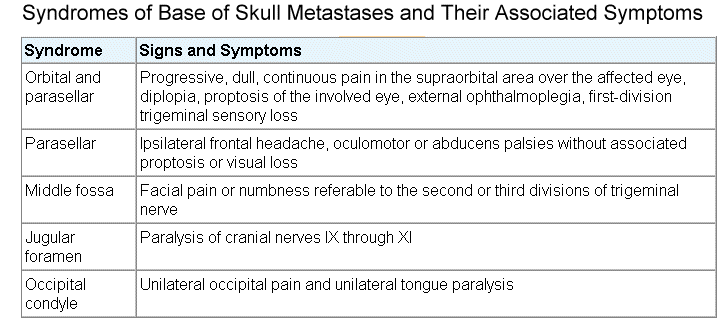
Metastases to the skull base usually fall within the province of the otolaryngological surgeon or oncologist. Cancers of the breast, lung, and prostate have a propensity to produce skull-base metastases, which can entrap the cranial nerves and vessels at their exit foramina. Greenberg has identified five clinical syndromes: orbital, parasellar, middle fossa, jugular foramen, and occipital condyle. Skull-base metastases most commonly involve the middle cranial fossa, in the area of Gasserian ganglion. Other sites include the jugular foramen, affecting cranial nerves IX, X, and XI to produce posterior auricular pain; occipital condyle, causing ipsilateral hypoglossal palsy and occipital pain; parasellar region, exhibiting as unilateral frontal sensory loss, headaches, and ophthalmoplegia; and orbit, presenting as proptosis and external ophthalmoplegia. Often the cancer appears as a thin skin of tumor cells, termed en plaque. Owing to their insidious subacute onset, skull-based metastases are difficult to identify on MRI or CT where masses may coalesce with changes of prior irradiation or surgical extirpation. Often the T2-weighted changes in contiguous bone after radiotherapy may be confused with tumor. Radionucleotide bone scans with careful evaluation of the skull may improve diagnosis. Treatment depends on the nature of the underlying tumor and is typically confined to irradiation as previously described. |
|
The presence of cranial neuropathy in cancer patients most commonly indicates the presence of meningeal carcinoma, tumor involvement of the bones of the cranial base with encroachment of neural foramina, or rarely, brainstem metastasis. Chemotherapy-induced cranial neuropathy is rare. Vincristine can cause cranial nerve palsy; extraocular eye movement abnormalities are most common. Intraventricular administration of drugs and of biologic response-modifying agents can cause transient cranial nerve palsy, often due to abnormalities in CSF circulation.
Clinical Manifestations and Differential Diagnosis
In both meningeal carcinoma and drug-induced cranial nerve palsy, the seventh (facial) cranial nerve is most commonly affected. Unfortunately, there is little to differentiate the clinical manifestations of drug-induced nerve palsy from those of meningeal carcinoma. Both conditions can involve several cranial nerves, be indolent in onset, and demonstrate spontaneous resolution. Therefore examination of the CSF is critical in the care of these patients. In addition to meningeal carcinoma, tumor encroachment on neural foramina at the skull base from bone metastasis can cause cranial nerve palsy, as can small intraparenchymal lesions (tumor, infarct, or hemorrhage), although such lesions are rare.
Diagnostic Evaluation
In the care of patients with cranial nerve palsy, several examinations of the CSF often are needed to determine whether tumor cells are present. Evaluation of the bones of the skull base with thin-section CT helps determine the presence of metastatic bone lesions. MRI of the brainstem is performed to evaluate for intraparenchymal lesions, although in most cases, other brainstem signs are present, in which case, this test should be part of the initial evaluation.