| From the RTOG 97-13 skin protocol; During a course of radiation temporary
skin reactions are common and vary from mild erythema to brisk, moist desquamation. The
severity of skin toxicity is dependent on volume of tissue treated, total daily dose,
fractionation, dose distribution, and certain individual factors. These reactions
are commonly treated with local measures (i.e. creams, gels etc.) and occasionally
may require a treatment break. Skin reactions associated with radiation therapy may impose
significant discomfort and may interfere with the patient’s daily living activities.
A recent review of the literature for
radiation induced skin toxicity clearly demonstrated that no standard of care for the
prevention or intervention of radiation-induced skin toxicity exists, and that
intervention is primarily based on the clinician clinical experience. A random survey of
RTOG institutions revealed that 50% of the RTOG institutions surveyed utilize Aloe Vera
Gel as the treatment of choice for mild to moderate radiation induced dermatitis, while
the remainder of institutions utilized Aquaphor, or other products such as Carasyn gel,
Lanolin etc. Although much attention has been given to how radiation therapy affects the
skin, until recently little research has been performed in identifying and standardizing
clinical intervention. Clinically, the emphasis has been on teaching self care to minimize
skin trauma, irritation, and/or infection.
A study in press by the North Central
Cancer Treatment Group reported that Aloe Vera Gel did not protect against
radiation-induced dermatitis when used prophylactically in women undergoing breast
irradiation. Another study performed in 1990 at Rush-Presbyterian-St. Luke’s
Medical Center, utilizing the product “Natural Care Gel,” with major ingredients
Aloe Vera, and D-panthenol, for women undergoing breast irradiation, demonstrated little
change in erythema or desquamation but did provide relief from burning and itching.
Biafine, a wound healing product from
France, was approved in 1995 by the FDA for use in the U.S. It has reportedly been the
product of choice in France for radiation induced dermatitis for two decades. A
comparative study of Biafine performed in 1973, at the Regional Centre for Combating
Cancer, at Marseille Hospital, France, concluded that Biafine was twice as effective as
the best alternative treatment for preventing and treating radiation skin reactions.
Another study concluded that Biafine enhances the first stage of the healing process by
recruiting macrophages and acts on the production of granulation tissue.
Earlier trials testing such agents as
topical vitamin C and topical cortisone cream have demonstrated no discernible benefit in
preventing radiation induced dermatitis. There are many factors that influence the effects
of radiation: site, time-dose volume relationships, radiation type and energy, nutritional
status and individual patient factors (i.e. complexion.) Thus, it will be important
to control for these factors. In today's health care setting, the importance and
consideration of cost containment cannot be discounted. Thus, it is important to monitor
cost/benefit ratios due to the variance among product costs. At the present time, there is
no standard of care for decreasing or preventing radiation-induced dermatitis. Presently,
there are numerous products being used by radiation oncology departments in the U.S. Based
on the results of the NCCTG and Rush-Presbyterian studies and the introduction of a new
skin care product (claiming) efficiency in preventing radiation-induced dermatitis,
this study is designed to compare other products with Biafine in preventing
radiation-induced dermatitis. It is hypothesized that Biafine will be a prophylactic agent
for radiation dermatitis in women undergoing breast irradiation.
Phase II study assessing the
effectiveness of Biafine cream as a prophylactic agent for radiation-induced acute skin
toxicity to the breast in women undergoing radiotherapy with concomitant CMF chemotherapy
Szumacher E, Wighton A, Franssen E, Chow E, Tsao M, Ackerman I, Andersson L, Kim J,
Wojcicka A, Ung Y, Sixel K, Hayter C
International Journal of Radiation Oncology*Biology*Physics - 01 September 2001 (Vol. 51,
Issue 1, Pages 81-86)
Purpose: To assess the efficacy of
Biafine cream in preventing Grade 2 acute radiation dermatitis, according to the National
Cancer Institute of Canada skin radiation toxicity criteria in patients undergoing
concomitant adjuvant chemotherapy and radiotherapy to the breast.
Methods and Materials: Sixty patients participated in this study. Patients were treated
with a lumpectomy followed by concomitant chemotherapy and radiotherapy to the breast.
Biafine cream was applied daily, starting on the first day and ending 2 weeks
post-radiotherapy. Patients underwent weekly skin assessments throughout radiotherapy and
at 2 and 4 weeks after treatment. Outcome measures were assessed using a Skin Assessment
Questionnaire that was scored according to the National Cancer Institute of Canada skin
radiation toxicity criteria and a self-administered questionnaire that evaluated skin
symptoms.
Results: The maximum skin toxicity observed during the course of treatment was as follows:
less than Grade 2 toxicity, 15% (9 patients); Grade 2, 83% (50 patients); Grade 3, 2% (1
patient); Grade 4, 0% (0 patients). The majority of the radiation dermatitis was observed
after 3 weeks of radiotherapy.
Conclusion: The majority of patients who underwent concomitant chemo- and radiotherapy for
breast cancer developed Grade 2 radiation dermatitis with the use of Biafine cream.
However, no treatment delays or interruptions were observed because of skin toxicity.
NCIC acute toxicity criteria for
radiation dermatitis
Rating Symptom
0 None
1 Faint erythema or dry desquamation.
2 Moderate to brisk erythema. Patchy moist desquamation less than 1.5 cm
mostly confined to skin folds and creases. Moderate edema.
3 Confluent moist desquamation greater than 1.5 cm not confined to skin.
Pitting edema.
4 Skin necrosis or ulceration of full-thickness dermis may include
bleeding—not induced by minor trauma or abrasion.
Phase III randomized trial of
Calendula officinalis compared with trolamine for the prevention of acute dermatitis
during irradiation for breast cancer.
Pommier P, Gomez F, Sunyach MP, D'Hombres A, Carrie C, Montbarbon X. J Clin
Oncol. 2004 Apr 15;22(8):1447-53.
Department of Radiation Oncology, Centre Leon Berard, France.
PURPOSE: The effectiveness of nonsteroid topical agents for the prevention of acute
dermatitis during adjuvant radiotherapy for breast carcinoma has not been demonstrated.
The goal of this study was to compare the effectiveness of calendula (Pommade au Calendula
par Digestion; Boiron Ltd, Levallois-Perret, France) with that of trolamine (Biafine;
Genmedix Ltd, France), which is considered in many institutions to be the reference
topical agent. PATIENTS AND METHODS: Between July 1999 and June 2001, 254 patients who had
been operated on for breast cancer and who were to receive postoperative radiation therapy
were randomly allocated to application of either trolamine (128 patients) or calendula
(126 patients) on the irradiated fields after each session. The primary end point was the
occurrence of acute dermatitis of grade 2 or higher. Prognostic factors, including
treatment modalities and patient characteristics, were also investigated. Secondary end
points were the occurrence of pain, the quantity of topical agent used, and patient
satisfaction. RESULTS: The occurrence of acute dermatitis of grade 2 or higher was
significantly lower (41% v 63%; P <.001) with the use of calendula than with trolamine.
Moreover, patients receiving calendula had less frequent interruption of radiotherapy and
significantly reduced radiation-induced pain. Calendula was considered to be more
difficult to apply, but self-assessed satisfaction was greater. Body mass index and
adjuvant chemotherapy before radiotherapy after lumpectomy were significant prognostic
factors for acute dermatitis. CONCLUSION: Calendula is highly effective for the prevention
of acute dermatitis of grade 2 or higher and should be proposed for patients undergoing
postoperative irradiation for breast cancer.
Randomized phase III study comparing
Best Supportive Care to Biafine as a prophylactic agent for radiation-induced skin
toxicity for women undergoing breast irradiation: Radiation Therapy Oncology Group (RTOG)
97-13.
Fisher J, Scott C, Stevens R, Marconi B, Champion L, Freedman GM, Asrari F, Pilepich MV,
Gagnon JD, Wong G. Int J Radiat Oncol Biol Phys. 2000 Dec 1;48(5):1307-10.
Radiation Oncology Center, Oakwood Hospital and Medical Center, Dearborn, MI 48123, USA. fisherj@oakwood.org
Approximately 180,000 women will be diagnosed with breast cancer in 1999; a large
percentage of these women will receive radiation therapy. It is estimated that 87% of
these women will develop some degree of radiation-induced dermatitis, varying from mild to
brisk erythema or even moist desquamation. The severity of skin toxicity is dependent on
volume of tissue treated, total daily dose, fractionation schemes, dose distribution and
certain individual factors (1). Skin reactions associated with radiation therapy can
impose significant discomfort and interfere with the patient’s daily living
activities and quality of life. Although much attention has been given to how radiation
therapy affects the skin, until recently, little research has been performed in
identifying and standardizing clinical intervention. Clinically, the emphasis has been on
teaching self-care to minimize skin trauma, irritation and infection. A random survey of
Radiation Therapy Oncology Group (RTOG) institutions in 1995, revealed that 50% of the
RTOG institutions utilized Aloe Vera Gel as the treatment of choice. The remaining
institutions utilized Aquaphor (Biersdorf, Lindenhurst, NY) or other products (Carasyn
Gel, Lanolin, etc.).
A study completed in 1991 by the North Central Cancer Treatment Group (NCCTG), reported
that Aloe Vera Gel did not protect against radiation-induced dermatitis when used
prophylactically in women undergoing breast irradiation (2).
Biafine (Medix Pharmaceuticals, Tampa, Florida), a wound-healing product from France,
approved in 1995 by the FDA for use in the United States as a wound dressing emulsion, has
reportedly been the product of choice in France for radiation-induced dermatitis for two
decades. A comparative study of Biafine performed in 1973 at the Regional Center for
Combating Cancer, at Marseilles Hospital, France, concluded that Biafine was twice as
effective in preventing and treating radiation skin reactions (3). Another French study
concluded that Biafine enhanced the first stage of the healing process by recruiting
macrophages, which initiates the production of granulation tissue (4).
Presently, there are numerous products being used by radiation oncology departments in the
United States. Based on the results of the NCCTG study and the introduction of a new
skin-care product claiming efficacy in preventing radiation-induced dermatitis, this study
was designed to compare Biafine to best supportive care (BSC) in preventing
radiation-induced dermatitis.
PURPOSE: To determine if Biafine compared to Best Supportive Care (BSC) is effective in
minimizing or preventing radiation-induced dermatitis in women undergoing breast
irradiation. METHODS AND MATERIALS: Patients were randomized between Biafine (n = 83) vs.
BSC (n = 89). The institutions identified preference for BSC at the time of randomization.
A no-treatment arm was allowed (16% received no treatment). Patients were instructed to
apply randomized product three times a day, but not within 4 h of their daily RT session.
Application began following their first radiation treatment and continued 2 weeks
postradiation. Skin dermatitis was scored weekly utilizing the RTOG and ONS (Oncology
Nursing Society) skin toxicity scales, a weekly patient satisfaction and quality-of-life
questionnaire. RESULTS: Using the RTOG toxicity scale there was no overall difference for
maximum dermatitis during RT between Biafine and BSC (p = 0.77). There was no difference
in maximum toxicity by arm or breast size. There was an interaction between breast size
and toxicity, with large-breasted women exhibiting more toxicity. Large-breasted women
receiving Biafine were more likely to have no toxicity 6 weeks post RT. CONCLUSION: There
was no overall difference between BSC and Biafine in the prevention, time to, or duration
of radiation-induced dermatitis.
In the design of this trial it was estimated that skin toxicity would have
the following profile: 13% Grade 0; 52% Grade 1; 32% Grade 2 and 2% Grade 3; and 1% Grade
4 based on prior RTOG experience. The actual observed toxicity was 7%, 58%, 32%, 3%, and
0%. This trial was unable to support the prestudy hypothesis that Biafine was more
effective than BSC in preventing or minimizing radiation-induced dermatitis. There was no
overall difference between Biafine and BSC in prevention, time to, or duration of
radiation-induced dermatitis. There was a slight statistical benefit for large-breasted
women and in nonsmoking women for reduction in skin toxicity postradiation therapy with
Biafine. However, this trial was designed to find a prevention effect, not an intervention
effect. The timing of Biafine vs. BSC in a group of patients at greater risk to develop
increased severity of skin toxicity is currently being planned within the RTOG.
The patient population from this trial are comparable with the NCCTG trial: 11% (NCCTG)
and 10% (RTOG) pigmented patient population. This prevented investigating skin toxicity or
intervention by pigmentation.
A method for preventing or minimizing radiation-induced dermatitis in the breast
population remains unanswered. There is little scientific or clinical evidence that
Biafine is superior to other emollients. This could partly be due to problems with this
study’s methodology. The study was not blinded therefore allowing bias. The use of a
skin ruler to provide consistency for measuring and documenting skin toxicity might have
been beneficial. Eligibility criteria could have included a more at risk patient
population, i.e., post mastectomy patients or post chemotherapy patients. The inability to
address skin toxicity and analyze response for the pigmented skin population was a
disadvantage. Patients compliance in following the skin care regimen was not objectively
monitored with a patient diary. Last, the question of timing for clinical intervention
remains unanswered. Neither this trial or the NCCTG trial provided clear answers to this
pertinent question.
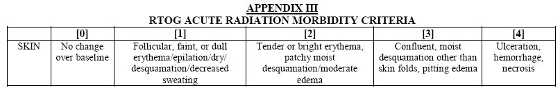
Skin toxicity is a side effect addressed daily in our practices. A Grade 2 skin toxicity
is described in the RTOG Acute Morbidity Scale as, “tender or bright erythema, patchy
moist desquamation/moderate edema.” Although not considered a major toxicity, 32% of
the women in this study developed a Grade 2 skin toxicity which was equated with a
meaningful decrease in their QOL. As professionals there remains a need to continue
investigating new products, techniques and novel approaches for minimizing or preventing
radiation-induced dermatitis in this patient population.
Topical corticosteroid therapy for
acute radiation dermatitis: a prospective, randomized, double-blind study.
Schmuth M, Wimmer MA, Hofer S, Sztankay A, Weinlich G, Linder DM, Elias PM, Fritsch PO,
Fritsch E. Br J Dermatol. 2002 Jun;146(6):983-91.
Department of Dermatology, University of Innsbruck, Austria. matthias.schmuth@uibk.ac.at
BACKGROUND: Radiation dermatitis is a common side-effect of radiation therapy, but there
is no current consensus about its appropriate therapy. OBJECTIVES: To compare treatment
with topical 0.1% methylprednisolone vs. 0.5% dexpanthenol in a cohort of patients
undergoing fractionated radiation therapy for breast cancer. METHODS: In a randomized,
double-blind design, treatment was initiated at the beginning of radiation therapy and
continued for 2 weeks after termination of radiation. Outcomes were compared by three
different measures: clinical (symptom score), functional (transepidermal water loss, TEWL)
and subjective (quality of life, QOL). RESULTS: In a preliminary cohort of untreated
patients undergoing radiation therapy, clinical signs and TEWL levels increased
progressively during radiation therapy, reaching highest values at 5 and 4 weeks,
respectively. Although neither topical treatment reduced the incidence of radiation
dermatitis, both delayed the emergence of greatest clinical and TEWL scores until
approximately 6 and 5 weeks, respectively. With topical corticosteroids, clinical symptoms
and TEWL were less pronounced than with dexpanthenol. Whereas general QOL improved after
completion of radiation therapy, skin-related QOL declined. However, the skin-related QOL
decline could be at least in part reversed by use of topical corticosteroid vs.
dexpanthenol-containing emollient. CONCLUSIONS: We provide evidence that prophylactic and
ongoing use of topical therapy with either topical corticosteroid or a
dexpanthenol-containing emollient ameliorates, but does not prevent radiation dermatitis.
Our data suggest, but do not prove, a benefit of a topical corticosteroid vs. a
dexpanthenol-containing emollient. Further controlled studies with larger cohorts will be
needed to determine optimal forms of topical therapy for radiation dermatitis.
The impact of skin washing with water
and soap during breast irradiation: a randomized study.
Roy I, Fortin A, Larochelle M. Radiother Oncol. 2001 Mar;58(3):333-9.
Department of Radiation Oncology, Centre Hospitalier Universitaire de Quebec, Pavillon
L'Hotel-Dieu de Quebec, 11, Cote-du-Palais, Quebec City, Quebec, Canada.
BACKGROUND: The effect of washing the irradiated skin during radiotherapy for breast
cancer is uncertain. The purpose of this study was to evaluate the impact of washing the
breast skin with water and soap during radiotherapy on the intensity of acute skin
toxicity. MATERIALS AND METHODS: Ninety-nine patients treated for breast cancer were
prospectively randomized prior to receiving radiotherapy to the breast into two groups:
(1), no washing was allowed during radiotherapy (49 patients); and (2), washing was
allowed with water and soap (50 patients). Acute toxicity was recorded according to the
Radiation Therapy Oncology Group (RTOG) acute skin toxicity scale for each patient every
week during radiotherapy and 1 month after the end of radiotherapy. Symptoms related to
skin toxicity were scored by visual analogue scales at the same time intervals. Other data
collected included sociodemographic data, characteristics related to the tumor and
previous treatments, radiation technique, necessity for a second simulation due to loss of
skin marks and treatment interruptions. RESULTS: In the non-washing group, the following
maximum acute toxicity scores were observed: grade 0, 2%; grade 1, 41%; grade 2, 57%;
grades 3 and 4, 0%. For the washing group, the scores were: grade 0, 0%; grade 1, 64%;
grade 2, 34%; grade 3, 2%; and grade 4, 0%. Moist desquamation was seen in 33% of
non-washing patients, but in only 14% of washing patients. The median scores of pain,
itching and burning of the treated skin were higher in the non-washing group, although
this was not statistically significant. In a multivariate analysis using logistic
regression, acute skin toxicity was associated with the patient's weight, concomitant
radiochemotherapy and hot spots on dosimetry, and there was a trend toward more acute skin
toxicity in the non-washing group. CONCLUSION: Washing the irradiated skin during the
course of radiotherapy for breast cancer is not associated with increased skin toxicity
and should not be discouraged.
Does aqueous or sucralfate cream
affect the severity of erythematous radiation skin reactions? A randomised controlled
trial.
Wells M, Macmillan M, Raab G, MacBride S, Bell N, MacKinnon K, MacDougall H, Samuel L,
Munro A. Radiother Oncol. 2004 Nov;73(2):153-62.
School of Nursing and Midwifery, University of Dundee, 11 Airlie Place, Dundee DD1 4HJ,
UK.
BACKGROUND AND PURPOSE: Evidence on which to base decisions about the management of
radiation skin reactions is lacking. The purpose of this study was to investigate whether
sucralfate or aqueous cream reduced acute skin toxicity during radiotherapy to the head
and neck, breast or anorectal area (phase A), and to evaluate the effect of hydrogels and
dry dressings on moist desquamation (phase B). This paper presents the results of phase A.
PATIENTS AND METHODS: Three hundred and fifty seven patients were randomised to apply
aqueous cream, sucralfate cream or no cream to the irradiated area from day one of radical
radiotherapy treatment. All patients were instructed to wash using unperfumed soap. Acute
skin toxicity was measured using a modified radiation therapy oncology group (RTOG) score,
reflectance spectrophotometry, patient diary card and dermatology life quality index
(DLQI). A cost minimisation approach was used to compare the costs of each skin care
approach. RESULTS: No consistent differences were found in the severity of skin reactions
or levels of discomfort suffered by patients in each of the randomised groups. Patients
with a higher body mass index, who smoked, received concomitant chemotherapy, boost or
bolus during treatment were more likely to develop skin reactions. CONCLUSIONS: There is
no evidence to support the prophylactic application of either of the creams tested for the
prevention of radiation skin reactions. Our results show that it is possible to predict
which patients are at greatest risk of skin reactions. We suggest that known risk factors
should be incorporated into future study protocols.
The standardization of radiation skin
care in British Columbia: a collaborative approach.
Nystedt KE, Hill JE, Mitchell AM, Goodwin F, Rowe LA, Wong FL, Kind AL. Oncol Nurs Forum.
2005 Nov 3;32(6):1199-205
Radiation Therapy at Vancouver Island Centre in the British Columbia.
knystedt@bccancer.bc.ca
PURPOSE/OBJECTIVES: To develop evidence-based practice guidelines for and standardize the
care of radiation skin reactions. DATA SOURCES: Peer-reviewed scientific journals and
texts and a survey of the guidelines in use at leading cancer treatment facilities in
Canada, the United States, the United Kingdom, and Australia. DATA SYNTHESIS: A formal
reference document with recommended guidelines was developed. Consensus was obtained from
all relevant disciplines, and the guidelines were implemented successfully into practice.
CONCLUSIONS: The document introduced a major change in practice from the maintenance of a
dry radiation treatment area to the promotion of skin cleanliness and hydration, as well
as the adoption of the principles of moist wound healing. Annual review indicated that
dissemination of (94%) and compliance with (78%) the guidelines were good. IMPLICATIONS
FOR NURSING: The process to develop, obtain consensus for, and implement evidence-based
practice guidelines was an exemplary demonstration of teamwork and interdisciplinary
collaboration.
Prevention and treatment of acute
radiation dermatitis: a literature review.
Wickline MM. Oncol Nurs Forum. 2004 Mar-Apr;31(2):237-47
Seattle Cancer Care Alliance, Seattle, Washington, USA. mihkai@u.washington.edu
PURPOSE/OBJECTIVES: To review historical and current research data on prevention and
treatment of acute radiation dermatitis. DATA SOURCES: 18 research trials and 1 case
report published from 1967-2001 and 1 unpublished research trial from 1972. DATA
SYNTHESIS: Washing the skin with mild soap and water and the hair with mild shampoo is
safe during radiation therapy. Biafine (Medix Pharmaceuticals, Inc., Largo, FL), chamomile
cream, almond ointment, topical vitamin C, and gentian violet have not been proven
effective and should not be used. Transparent, hydrocolloid, and hydrogel dressings have
been beneficial, as have sucralfate cream and corticosteroid cream. Aloe vera may be
beneficial and is not harmful. CONCLUSIONS: The existing scientific data are lacking in
quantity and quality. The current body of evidence is unable to provide clinicians with
comprehensive guidelines for prevention and management of acute radiation dermatitis.
IMPLICATIONS FOR NURSING: Nurse clinicians and nurse scientists must partner to conduct
further research to add to the limited resources about the prevention and management of
acute radiation dermatitis and develop comprehensive evidence-based clinical practice
guidelines. |