| Treatment Basal Cell Carcinoma Mohs' Micrographic Surgery Habif: Clinical Dermatology, 3rd ed., Copyright © 1996 Mosby-Year Book, Inc. |
|
| Treatment Basal Cell Carcinoma Mohs' Micrographic Surgery Habif: Clinical Dermatology, 3rd ed., Copyright © 1996 Mosby-Year Book, Inc. |
|
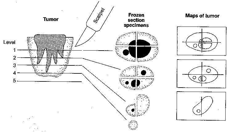
Mohs' Micrographic Surgery Rather than increase in a sphere-shaped mass, certain skin tumors, such as basal cell epithelioma, transmit random, fingerlike projections into the surrounding connective tissue. These tumor strands may go undetected with standard desiccation and curettage or excision techniques, resulting in recurrence. In the past, multiple procedures were performed on unfortunate patients who, after a series of unsuccessful procedures, acquired a diffuse, poorly defined mass of substantial proportions. In 1941, Frederick Mohs described a microscopically guided method of tracing and removing basal cell carcinomas. Since then the technique has been used to treat many contiguously spreading skin cancers. [39] The procedure has been modified and is usually performed on fresh tissue in 1 day on an outpatient basis. Tissue is removed in thin layers, and all margins of the specimen are mapped to determine whether tumor remains. Cure rates are very high. The technique is tissue sparing: The tumor is precisely identified and maximum amounts of normal skin can be retained. TECHNIQUE--MOHS' MICROGRAPHIC SURGERY The clinically apparent area is removed with a curet . In the past, a chemical fixative--zinc chloride paste--was applied and allowed to penetrate the tissue. Paste application resulted in the use of the now outdated term, Mohs' chemosurgery. Currently, the fixation step is omitted, thus the designation fresh tissue technique. A thin, horizontal layer of tissue is removed with a scalpel and divided into more convenient smaller specimens for frozen section. Two adjacent edges of tissue are dyed red and blue to provide spatial orientation. A diagram of the section is prepared, and the number and color coding is indicated on the map. Specimens are mounted in a cryostat and then sectioned. Cut sections are stained and microscopically examined. The location of the tumor cells is indicated on a map and the above steps are repeated only in areas with tumor until a cancer-free plane is reached. The defect created by the fresh tissue technique can heal by secondary intention or can be closed primarily. Flaps were found to be preferable to skin grafts for facial repair, with forehead and nasolabial flaps particularly useful for the nose. Cure rates of 94% to 99% have been achieved The advantages of the microscopically controlled technique are that it preserves maximum amounts of normal tissue around the cancer, and it provides great reliability in determining adequate margins of excision. The disadvantage is that it is time-consuming, requiring hours or days to perform. |
|
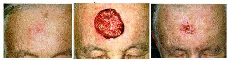
Mohs' micrographic surgery technique.A, Sclerosing basal cell carcinoma. A small nodule is surrounded by an ill-defined erythematous area of induration.B, Mohs' micrographic surgery reveals the extent of the tumor shown, which clinically appeared to be rather small.C, Six weeks after Mohs' micrographic surgery. The defect is healing by secondary intention. |