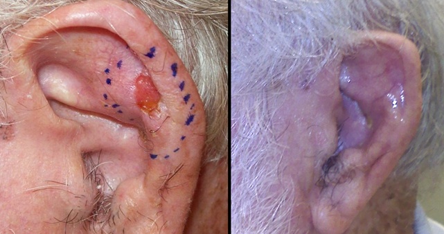
| Necrosis following radiotherapy for
carcinoma of the pinna.
Hayter CR, Lee KH, Groome PA, Brundage MD. Int J Radiat Oncol Biol Phys
1996 Dec 1;36(5):1033-7
Radiation Oncology Research Unit, Queen's University, Kingston General Hospital, Ontario,
Canada.
Radiation therapy is often the preferred modality of treatment for carcinoma of the pinna
because it avoids the cosmetic defect of surgery. However, radiation oncologists are
sometimes reluctant to irradiate the ear because of the risk of subsequent necrosis. The
goal of this study was to establish the long-term disease control and necrosis rates
following irradiation of the external ear. METHODS AND MATERIALS: A retrospective analysis
was undertaken of 138 courses of curative radiotherapy given to 128 patients for
biopsy-proven basal (70 courses), squamous (62 courses), or mixed (6 courses) tumors of
the pinna between January 1, 1982, and December 31, 1991, at the Kingston Regional Cancer
Center. RESULTS: The median age of the patients was 73 (range 43-94) and the median size
of the tumors was 12 mm (range 3-50 mm). Treatment was given using orthovoltage X rays
(79) or electrons (59). The most common dose prescription was 35 Gy/5 fractions; total
doses ranged from 17.50 to 64 Gy. The median follow-up is 58 months (range 6-149). The actuarial 5-year local control rate is 93%; the actuarial necrosis rate
at 5 years is 13%. Most necroses healed with conservative management; only two
patients required surgery for necrosis. We analyzed the following factors as possible
predictors of radiation necrosis: patient age, size of lesion, histology, fraction size,
total dose, overall time, and beam energy. Only daily fraction
sizes > 6 Gy (p = 0.0093) and treatment times < 5 days (p = 0.0053) were
significantly associated with an increased risk of necrosis.CONCLUSION: To
reduce the risk of necrosis, radiation therapy for external ear cancer should be given
using protracted fractionation.
Megavoltage electron beam therapy in the treatment of
basal and squamous cell carcinomata of the pinna.
Hunter RD, Pereira DT, Pointon RC. Clin Radiol 1982 May 3;33(3):341-5
Kilovoltage X-ray therapy has considerable limitations when trying to obtain good
functional results in patients with skin carcinomas arising on the pinna. Megavoltage
electron beams with their better quality of radiation and homogeneous dose distribution
have been recognised to have theoretical advantages. Forty-three patients with basal and
squamous cell carcinomata arising on the pinna were treated radically using a 10 MeV
electron beam. The technique and dosage are described and discussed. Primary cancer
control with retention of the pinna was achieved in 34 patients. Salvage pinnectomy was
performed in four patients for recurrence and one patient for radiation necrosis. Two
patients with large primary tumours failed to resolve and died of their disease. The
advantages for the patient of the policy of primary radical electron mean therapy are
discussed.
Irradiation of the pinna with superficial kilovoltage
radiotherapy.
Lim JT. Clin Oncol (R Coll Radiol) 1992 Jul;4(4):236-9
Mersey Regional Oncology Centre, Bebington, Wirral, UK.
Excellent local control can be achieved by treating superficial carcinomas of the skin
with superficial kilovoltage radiotherapy. The results of this study support the use of
100-140 KV photons to treat carcinomas of the skin of or around the pinna. This achieves a local control rate of 97% and the radionecrosis rate of
10% could be halved if all patients were treated with 3500 cGy in 5 fractions over one
week, or to a maximum area of 5 cm2 of irradiated pinna. An audit of the
running cost reveals that this method of treating superficial carcinomas is highly cost
effective and can be recommended for all suitable patients.
Treatment results and patterns of failure in 646
patients with carcinoma of the eyelids, pinna, and nose.
Petrovich Z, Kuisk H, Langholz B, Astrahan M, Luxton G, Chak L, Rice D.
Department of Radiation Oncology, University of Southern California School of Medicine,
Los Angeles 90033. Am J Surg 1987 Oct;154(4):447-50
From 1956 to 1978, 646 patients were treated with radiotherapy for carcinoma of the nose
(350 patients, 54 percent), eyelids (159 patients, 25 percent), pinna (93 patients, 14
percent), and skin adjacent to the lip (44 patients, 7 percent). The histologic
distribution was 72 percent basal cell carcinoma, 18 percent squamous cell carcinoma, and
10 percent mixed basal and squamous cell features. Tumors less than 2 cm in diameter were
found in 602 patients (93 percent), whereas 44 patients (7 percent) had larger tumors.
Tumor involvement of cartilage and bone was seen in 23 patients at the time of diagnosis. The 5, 10, and 20 year control rates were 99 percent, 98 percent, and
98 percent, respectively, for 502 tumors less than 2 cm in diameter. This compared
favorably with control rates of 92 percent at 5 years and 79 percent at 10 years for
tumors from 2 to 5 cm in diameter and 60 percent at 5 years and 53 percent at 8 years for
12 patients with massive tumors (p less than 0.0001). The histologic
characteristics of the lesion had a strong influence on tumor control (p less than 0.02).
Of the patients with cartilage or bone invasion, tumor was controlled in 19 (83 percent).
Of these 19 patients, 11 had no evidence of disease for 3 years or more. Of all 646
patients treated, failure was seen in 60 (9 percent). It correlated well with the size of
the lesion, being 7 percent for tumors of less than 2 cm and 50 percent for tumors of
greater than 5 cm. Of the 60 patients in whom treatment failed, 48 (80 percent) had prior
definitive therapy. Radiotherapy was an efficient modality to control operative failures;
however, it was not as efficient at control in patients in whom previous radiotherapy
failed. Operation was the treatment of choice to salvage patients in whom radiotherapy
failed. Of the patients in whom retreatment failed, 10 were known to have died from skin
cancer, and an additional 6 patients were presumed to have died from the cancer. This study has demonstrated a good control rate and good cosmetic
results for small tumors of the eyelids, pinna, and nose. In addition, a good control rate
was obtained in patients with cartilage and bone involvement. Treatment of
massive tumors should involve planned operative resection with adjuvant radiotherapy.
Results of radiotherapy for epithelial skin cancer of
the pinna: the Princess Margaret Hospital experience, 1982-1993.
Silva JJ, Tsang RW, Panzarella T, Levin W, Wells W. Int J Radiat Oncol Biol
Phys 2000 May 1;47(2):451-9
Department of Radiation Oncology, Princess Margaret Hospital, University Health Network,
University of Toronto,
PURPOSE: To assess the treatment outcome, late toxicity, and prognostic factors for
radiotherapy (RT) of carcinoma of the pinna. METHODS AND MATERIALS: The charts of 313
patients treated between 01/82 and 12/93 were retrospectively reviewed. There were 334
lesions treated: 201 basal cell carcinoma (BCC), 122 squamous cell carcinoma (SCC), and 11
basosquamous carcinoma. RT was most commonly given by orthovoltage X-rays (278 lesions) or
electrons (39 lesions). The most frequently used dose prescriptions were 35 Gy in 5
fractions (123 treatments with median field size = 4.9 cm(2)), 42. 5-45 Gy in 10 fractions
(67 treatments with median field size = 10.5 cm(2)), and 50-65 Gy in 20-30 fractions (42
treatments with median field size = 81 cm(2)).2 cm. RESUL TS: The actuarial 2- and 5-year local control rates were 86.6% and 79.2 %.
Multivariate analysis revealed two factors to be statistically signi ficant for increased
local failure: tumor size > 2 cm (hazard ratio [HR] = 2.66, 95% confidence interval
[CI] = 1.16-6.08), and a low biological effective dose (BED) (for each decrease of 5 BED
units, HR = 1.76, 95% CI = 1.07-2.88). The 5-year actuarial rate of significant Grade 4
late toxicity was 7.3%. Factors statistically significant for this endpoint on univariate
analysis were tumor size (p = 0.035), T-stage (p = 0.02), field size (p = 0.05), fraction
size (p = 0.003), and BED (p = 0.05). CONCLUSIONS: RT is an eff ctive treatment option for
epithelial skin cancer of the pinna. Large t umor size and low BED were independently
statistically significantly ass ociated with increased local failure. Dose-fractionation schedules usin g fraction sizes < 4 Gy may reduce
the risk of necrosis and ulceration, particularly for field sizes > 5 cm2. |

