seminoma_port.jpg (30983 bytes) 
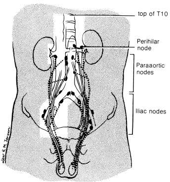
Radiation Therapy Techniques (from Perez)

Patients with stage I seminoma should receive megavoltage irradiation to the paraaortic and ipsilateral pelvic nodes (Fig on left). The treatment volume should include the superior plate of the T10 vertebra and should extend inferiorly to the top of the obturator foramina. It is unnecessary to include the scar in the inguinal region. The lateral borders should include the paraaortic lymph nodes, as visualized by lymphogram, and the ipsilateral renal hilum. The field is usually 10 to 12 cm wide except at the hila, where it may be wider. The ipsilateral iliac and pelvic nodes should be encompassed by a shaped field with 2-cm margins on the visualized nodes. Testicular shielding should be used if the patient wishes to preserve fertility.

The irradiation technique used is an anterior and posterior parallel opposed pair, using megavoltage equipment. Both fields should be treated daily, 5 days per week, Monday to Friday. It was agreed at the 1989 Consensus Conference that the recommended dose prescription should be 25 Gy given in 20 fractions. It is common practice in the United States to deliver about 25 Gy in 1.6- to 1.8-Gy fractions. Many centers commonly use higher doses, but all available data suggest that higher doses are unnecessary for sterilizing microscopic retroperitoneal disease

For stage II disease, the volume irradiated and the dose used are modified according to the bulk of the retroperitoneal disease. For stage IIA, no modification of the volume or dose of irradiation is necessary. For stage IIB disease (2 to 5 cm diameter), the field width should be appropriately widened to encompass the mass as visualized on CT or lymphography, with a margin of 2 cm . For masses with transverse diameters greater than 4 cm, the total irradiation dose is increased to 35 Gy. The first 25 Gy is delivered to an initial volume, and a boost of 10 Gy in five to eight fractions is given to a reduced field that encompasses the node mass with an adequate margin if the retroperitoneal mass exceeds 4 cm in diameter. Some radiation oncologists treat the contralateral and the ipsilateral pelvic nodes. This treatment is prescribed because of the risk of retrograde spread into the contralateral nodes from a relatively large retroperitoneal mass, particularly if it is low lying. However, no data exist to estimate the risk of contralateral pelvic nodal relapse in this circumstance. If bilateral pelvic nodal irradiation is used, a central pelvic shield should be employed.

In the rare event that mediastinal and supraclavicular irradiation is required for isolated progression of disease in those sites after chemotherapy, a dose of 25 Gy is given in fractions of 1.75 to 2 Gy. The portal arrangement should be that of an anterior and posterior opposed pair of fields to the mediastinum, wide enough to encompass the visible disease with a margin of 1 to 1.5 cm. The supraclavicular fossae usually can be adequately treated with a single anterior field.