 |
Ultrasound Mapping is the first step |
|
Ultrasound Mapping is the first step |
The patient has an ultrasound performed. The image shows a cross section of the gland and images are obtained every 5mm. The images appear on a screen with a grid(like a checkerboard) that divides the gland into 5mm boxes. The images with the grid are then entered into a computer that then determines which boxes to places seeds into. The computer calculates the strength and number of seeds necessary so that an even dose of radiation is spread throughout the gland. |
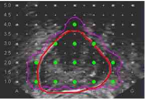 |
Ultrasound Image with grid lines in white, seed locations in green, prostate in red and radioactive field in purple |
![]()
![]()