Renal-Cell Carcinoma
Herbert T. Cohen, M.D., and Francis J. McGovern, M.D. , NEJM 2005;353:2477
In the United States, renal cancer is the 7th leading malignant
condition among men and the 12th among women, accounting for 2.6 percent of all cancers.
About 2 percent of cases of renal cancer are associated with inherited syndromes. In the
United States, 36,160 new cases of renal cancer are predicted to occur in 2005, many of
which are being discovered earlier because of the widespread availability of radiographic
testing. Nevertheless, 12,660 deaths from the disease are predicted to occur in 2005.
Renal-cell carcinomas arise from the
renal epithelium and account for about 85 percent of renal cancers. A quarter of
the patients present with advanced disease, including locally invasive or metastatic
renal-cell carcinoma. Moreover, a third of the patients who undergo resection of localized
disease will have a recurrence. Median survival for patients with metastatic disease is
about 13 months. Thus, there is a great need for more effective surgical and medical
therapies.
he classic presentation of renal-cell carcinoma
includes the triad of flank pain, hematuria, and a palpable abdominal mass. Few patients
now present in this manner. Roughly half the cases are now detected
because a renal mass is incidentally identified on radiographic examination. Other
common presenting features may be nonspecific, such as fatigue, weight loss, or anemia.
Risk factors for renal-cell carcinoma include smoking, obesity, and hypertension, as well
as acquired cystic kidney disease associated with end-stage renal disease. A 1.6:1.0 male
predominance exists,1 and the peak incidence is in the sixth and seventh decades. Gross or
microscopic hematuria is an important clinical clue to the diagnosis of renal-cell
carcinoma; thus, hematuria should be evaluated promptly by a computed tomographic (CT)
scan of the genitourinary tract and, in patients older than 40 years of age, by cystoscopy
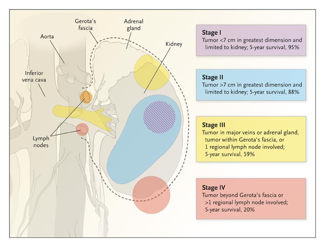
to rule out bladder cancer. Prognosis is closely related to the stage of disease (Figure 1).
Papillary Renal-Cell Carcinoma
Sporadic papillary renal-cell carcinoma has a five-year survival
rate approaching 90 percent and a striking 5:1 male predominance. Localized
papillary renal-cell carcinoma metastasizes less frequently than clear-cell renal-cell
carcinoma. However, the survival rate for metastatic papillary renal-cell carcinoma is
probably worse than that for clear-cell renal-cell carcinoma. The risk of both types is
particularly increased among patients with end-stage renal disease. Chromosome 7, which
harbors the MET proto-oncogene, is duplicated in 75 percent of sporadic papillary cases.
There are two subtypes of papillary renal-cell carcinoma. Type 1 tumors are papillary
lesions covered by small cells with pale cytoplasm and small oval nuclei with indistinct
nucleoli, and type 2 tumors are papillary lesions covered by large cells with abundant
eosinophilic cytoplasm. Type 2 cells are typified by pseudostratification and large,
spherical nuclei with distinct nucleoli. Type 2 tumors are genetically more heterogeneous,
have a poorer prognosis, and may arise from type 1 tumors.
Oncocytoma and Chromophobe Renal-Cell Carcinoma
Oncocytomas, which are benign, account for about 4 percent of nephrectomies performed
because renal-cell carcinoma is suspected. The chromophobe variant of renal-cell carcinoma
also accounts for 4 percent of all cases of renal-cell carcinoma and may have a benign
course after surgery, provided that the tumor stage and grade are favorable. Oncocytoma is
thought to originate from type A intercalated cells of the collecting duct, whereas
chromophobe renal-cell carcinoma is thought to originate from type B intercalated cells.
Collecting-Duct Renal-Cell Carcinoma
Collecting-duct renal-cell carcinoma accounts for less than 1 percent of all cases of
renal-cell carcinoma and is typically an aggressive tumor. Medullary carcinoma of the
kidney, which may be a variant of the collecting-duct type, is associated with sickle cell
trait or disease. The collecting-duct form may be most similar to transitional-cell
carcinoma of the urothelium.
Management of Sporadic and Hereditary
Renal-Cell Carcinoma
An enhancing renal mass on a CT scan obtained after the administration of contrast
material is a strong clue that renal cancer is present. A staging workup should be
performed before treatment is initiated. Multiple enhancing lesions, or a family history
of renal-cell carcinoma, particularly in persons younger than 50 years of age, suggests a
hereditary predisposition to the disease. Von Hippel–Lindau disease, hereditary
leiomyomatosis and renal-cell cancer, and the Birt–Hogg–Dub� syndrome all have
extrarenal manifestations, whereas familial clear-cell renal cancer and hereditary
papillary renal carcinoma do not. Thus, a careful physical examination including
ophthalmologic, neurologic, and dermatologic evaluation may be helpful. CT scanning or
magnetic resonance imaging (MRI) of the abdomen and pelvis may reveal uterine tumors in
patients with hereditary leiomyomatosis and renal-cell cancer or renal cysts or pancreatic
or adrenal involvement in patients with von Hippel–Lindau disease.
Patients with hereditary renal-cell carcinoma should be closely monitored. CT before and
after the administration of contrast material is the best test for detection and
assessment of renal masses, with gadolinium-enhanced MRI as an alternative. Such studies
can be performed at intervals ranging from every three to six months to every two to three
years, depending on the size of the lesions and the type of syndrome. Larger masses
require more frequent evaluation. Because small masses are usually of low grade, they can
be observed until they reach 3 cm, at which time they should be removed. However, tumors
caused by hereditary leiomyomatosis and renal-cell cancer should be excised immediately
because of their aggressive nature. Patients with von Hippel–Lindau disease should
undergo MRI studies of the brain and spinal cord to screen for hemangioblastoma. A family
pedigree should be generated, and family members at risk should be encouraged to seek
medical attention. Testing is available for the VHL, MET, FH, and BHD genes. One goal of
such testing is to free unaffected family members from continued cancer screening.
Organizations such as the VHL Family Alliance (www.vhl.org) are a vital resource for
patients, families, physicians, and researchers.
Prognosis
Defining the prognosis of renal-cell carcinoma is important for both therapeutic
decision-making and counseling patients. For metastatic renal-cell carcinoma, poor
prognostic factors include a low Karnofsky performance-status score (a standard way of
measuring functional impairment in patients with cancer), a high level of serum lactate
dehydrogenase, a low hemoglobin level, and a high corrected level of serum calcium. The
University of California, Los Angeles, Integrated Staging System was developed to evaluate
the prognosis at diagnosis and in the presence of metastatic disease; it includes
tumor–node–metastasis (TNM) staging, the patient's score on the Eastern
Cooperative Oncology Group performance-status scale (another measure of functional
impairment in patients with cancer), and the Fuhrman nuclear grade, which assesses
histologic features of the tumor.65 This system has been used successfully in more than
4000 patients at eight international centers.
Surgical Treatment
Radical Nephrectomy
Surgical excision is the primary treatment for renal-cell carcinoma.
Radical nephrectomy, which includes removal of the kidney en bloc with Gerota's fascia,
the ipsilateral adrenal gland, and regional lymph nodes, has been the standard therapy,
although more limited approaches are being explored. The surgical approach is determined
by the size and location of the tumor within the kidney, the TNM stage, and any special
anatomical considerations.
Staging and evaluation for the presence of metastases, including a careful history-taking
and physical examination, should be completed before surgery. Routine laboratory studies
should include measurement of the hematocrit and serum levels of creatinine, calcium, and
alkaline phosphatase and a urinalysis for proteinuria. Imaging studies, such as
radiographs of the chest, CT of the abdomen and pelvis, and in some cases, MRI evaluation
of the renal vein and inferior vena cava, CT of the chest or head, or bone scanning may be
needed. The frequency of follow-up after surgery depends on the stage of the tumor.
Surgery for Metastatic Disease
Nephrectomy may be warranted, even in the presence of metastatic disease. The combination of interferon alfa and nephrectomy is superior to
interferon alfa alone, offering a survival advantage of 3 to 10 months. Surgical
excision of a solitary metastasis in patients with advanced renal-cell carcinoma is
recommended in many cases, but this approach has not yet been proved to be effective in
prolonging survival.
Nephron-Sparing Partial Nephrectomy
Nephron-sparing partial nephrectomy has gained acceptance for treating
tumors less than 4 cm in diameter. Other indications for partial nephrectomy may
include a solitary kidney, bilateral renal masses, or renal insufficiency, as well as the
presence of hypertension, diabetes, or hereditary renal-cell carcinoma syndromes. Results achieved with nephron-sparing surgery are similar to those with
radical nephrectomy, but a disadvantage is a rate of local recurrence of 3 to 6 percent.
Laparoscopic Nephrectomy
First reported in 1991, laparoscopic nephrectomy has accelerated the evolution toward
minimally invasive surgical management of renal-cell carcinoma. The benefits of the
laparoscopic approach include decreased postoperative pain, a shorter hospitalization, and
a quicker recovery. The laparoscopic approach has been used for both radical nephrectomy
and partial nephrectomy. The laparoscopic partial nephrectomy, however, is a technically
demanding procedure with the potential for increased perioperative complications.
Percutaneous Ablative Approaches
The most recent evolution in the surgical management of small tumors has been percutaneous
thermal ablative techniques that use radiofrequency heat ablation or cryoablation to
destroy tumor cells. A needle probe is advanced through the skin and directed into the
tumor under image guidance. Although early results of radiofrequency
ablation and cryoablation are encouraging, larger trials with long-term follow-up are
needed. The rates of complications appear to be low, but reported adverse events
include intraoperative and postoperative hemorrhage, urinary leakage, and injury to
adjacent structures. Because identification of the type of renal-cell carcinoma is
important, a core biopsy of the renal mass should be performed as part of the procedure.
Ideal candidates for minimally invasive percutaneous ablative therapy are patients with
tumors less than 3 cm in diameter who have serious coexisting conditions and for whom
standard approaches would pose substantial risks. Patients with multifocal tumors may also
benefit from minimally invasive percutaneous procedures. High-frequency focused ultrasound
applied externally to the body is being studied as another potential minimally invasive
therapy.
Medical Treatment
Medical therapies are generally offered for locally advanced or metastatic renal-cell
carcinoma, and much of the clinical experience with this approach is in patients with the
clear-cell type. Because response rates are low, the need to identify new therapeutic
agents is great.
Chemotherapy
Rates of response to chemotherapy alone are low (roughly 4 to 6
percent). Drug resistance may be related to the expression of the multidrug
resistance transporter in proximal-tubule cells — the cells from which clear-cell and
papillary renal-cell carcinoma may originate. Chemotherapy may be more efficacious for
advanced non–clear-cell renal-cell carcinoma, particularly the collecting-duct type.
A phase 2 trial of carboplatin and paclitaxel for the collecting-duct form of the disease
is under way.
Immunomodulatory Therapies
The value of immunomodulatory therapy for clear-cell renal-cell carcinoma is supported by
reports of occasional spontaneous tumor regression, infrequent complete regression of
metastatic disease with cytokine therapies, and promising early results with allogeneic
stem-cell transplantation and tumor vaccines. The goal of immunomodulatory therapy is to
boost either tumor antigenicity or host surveillance. Unique tumor antigens may also be
inducible in renal-cell carcinoma.
Interferon Alfa
About 14 percent of cases of metastatic clear-cell renal carcinoma
respond to interferon alfa alone. Various doses and routes have been used. The
median duration of response is six months and rarely exceeds two
years. Because the side effects of the drug are not onerous, it appears to be a
good choice to use in combination with other agents in experimental approaches.
Interleukin-2
High-dose interleukin-2 is the standard therapy for advanced
renal-cell carcinoma and is the only regimen for this disease approved by the Food and
Drug Administration. However, many patients with metastatic disease cannot take
high-dose interleukin-2, because it causes a capillary leak syndrome or because it is not
available in all treatment centers. High-dose interleukin-2 induces
responses in 21 percent of patients, as compared with only 13 percent of patients who
receive low-dose interleukin-2. The median duration of response
has been reported to be 54 months overall, and for those with a complete response,
the median duration of a response is yet to be reached. Interleukin-2 has also been used
in combination with other drugs, but it is unclear whether combined therapy achieves
better results than interleukin-2 alone. Thus, interleukin-2 is a highly effective therapy
for a subgroup of patients with metastatic disease. Identifying features predictive of a
response to interleukin-2 would represent a further advance, and efforts are being made to
identify patients with clear-cell renal carcinoma who would be likely to have a response
to interleukin-2 therapy on the basis of pathological characteristics and expression of
CA9.
Adjuvant Therapy
Given the high rate of recurrence of renal-cell carcinoma after nephrectomy, a follow-up
adjuvant approach would be desirable, especially for patients with high-risk, locally
advanced disease. However, conventional chemotherapy, interferon alfa, or even
interleukin-2 has not proved effective as an adjuvant therapy. Approaches currently being
tested include tumor vaccines and a monoclonal antibody directed against CA9.
Evolving Therapies
Stem-Cell Transplantation
Allogeneic stem-cell transplantation performed after the administration of a
non–marrow ablative regimen elicits a potent graft-versus-tumor effect and appears
promising for treating clear-cell renal-cell carcinoma. Protocols developed at the
National Institutes of Health have used myelosuppressive pretreatment, followed by an
infusion of donor CD34+ cells and T cells from an HLA-identical sibling. A course of
immunosuppressive agents, such as cyclosporine, is used to limit graft-versus-host disease
and is rapidly tapered. Twenty of the first 45 patients with
metastatic renal-cell carcinoma who underwent stem-cell transplantation had a response (44
percent). However, results in some other centers have been less promising. The
responses have correlated well with the development of graft-versus-host disease and with
the conversion of T-cell chimerism to full donor origin. One goal is to identify the tumor
epitopes that are initiating the graft-versus-tumor response to improve treatment
specificity. The two drawbacks to stem-cell transplantation have been severe
graft-versus-host disease, which can be life-threatening, and the need for a
haplotype-matched sibling donor. Prognosis is also an important guide to patient
selection, since responses take several months. The next generation of strategies for
stem-cell transplantation may include the use of tumor vaccines after transplantation as
well as the use of cytokine therapy to boost recipient or even donor immunity.
Tumor Vaccines
Tumor vaccines represent a potential means of enhancing host immunity. A promising
approach to the treatment of advanced clear-cell renal carcinoma uses autologous or donor
dendritic cells, which initiate a primary immune response by presenting antigen in the
context of costimulatory molecules. Dendritic cells can be pulsed with tumor protein, DNA,
or RNA; they can even be fused with tumor cells to present tumor antigens in a context
favorable for therapy. Such vaccines are generally well tolerated, but they will require
further optimization. Concomitant administration of cytokines may improve the response to
vaccines.
Target Antigens
A goal of stem-cell or vaccine therapies is to characterize the tumor antigens involved in
the immune response. One potential target is the G250 renal cancer antigen, which has been
identified as CA9. The CA9 gene is a target of HIF and so is overexpressed in VHL-related
clear-cell renal carcinoma, even in the earliest lesions of von Hippel–Lindau
disease. Thus, in cases of renal-cell carcinoma, a high proportion of CA9-positive cells
may be associated with a more favorable prognosis. As a transmembrane protein, CA9 may
also be a therapeutically useful tumor antigen.92,93 It will be important to identify
additional target antigens.
Therapies Targeting VEGF and TGF-{alpha} Pathways
Originally identified as regulated by VHL, VEGF and TGF-{alpha} are now promising
therapeutic targets in clear-cell renal carcinoma. The manner in which these molecules
interact with the cancer epithelium and surrounding vascular endothelium leads to tumor
progression. A combination of therapies based on rational targets such as these may
therefore be a powerful approach to advanced renal-cell carcinoma.
VEGF-Pathway Components as Molecular Targets
VEGF is overexpressed throughout clear-cell renal-cell carcinoma tissue and may be the
most important tumor angiogenic factor. A randomized phase 2 trial involving patients with
metastatic renal-cell carcinoma investigated the efficacy of bevacizumab, a humanized
VEGF-neutralizing antibody. This agent extended the interval before tumor progression to
4.8 months, as compared with 2.5 months for placebo. Bevacizumab therefore provided a key
"proof of principle" of the efficacy of anti-angiogenic therapy and may offer
additional benefit when given in combination with other drugs. Inhibitors of VEGF receptor
tyrosine kinase are being developed and tested. Indeed, the multitargeted kinase
inhibitors sunitinib and sorafenib have shown great promise in phase 2 and phase 3 trials,
with at least stabilization of disease in as many as 70 percent of patients with
cytokine-refractory disease.
TGF-{alpha}–Pathway Components as Molecular Targets
TGF-{alpha} is a potent growth factor for epithelial cells that acts through the epidermal
growth factor receptor (EGFR), which is a receptor tyrosine kinase. TGF-{alpha} is
overexpressed in the epithelium in clear-cell renal carcinoma and is a VHL target.
Overexpression of TGF-{alpha} is an early event in the pathogenesis of this disease.96
Furthermore, growth of renal cancer cells in culture is dependent on TGF-{alpha}. Thus,
the TGF-{alpha} pathway is a logical choice for therapeutic intervention.
Antibodies against EGFR are thought to bind EGFR and promote its down-regulation from the
cell surface. A fully human monoclonal antibody against human EGFR, called panitumumab
(ABX-EGF), has been evaluated in a phase 2 trial involving 88 patients with metastatic
renal-cell carcinoma.98 Only one patient had a complete response, and two had partial
responses — a disappointing result. Small-molecule inhibitors of the EGFR tyrosine
kinase are also being developed. The quinazolines gefitinib and erlotinib are now in phase
2 trials. In a phase 1 trial of erlotinib, just one patient with metastatic disease had a
complete response.100 Erlotinib is also being tested in combination with bevacizumab,
although encouraging initial results could not be confirmed in a randomized phase 2 trial.
Other Approaches
Temsirolimus (CCI-779), a selective inhibitor of the mammalian target of rapamycin, has
shown efficacy in a phase 2 trial of metastatic renal-cell carcinoma.101 Temsirolimus may
inhibit HIF as well. Partial responses were noted in 7 percent of patients, and minor
responses in 26 percent. The median survival rate was 15 months. The notable activity of
the drug in patients with poor prognostic features prompted a phase 3 trial. Other options
are being pursued, including agents targeting HIF.
Summary and Prospects for the Future
The poor prognosis of advanced renal-cell carcinoma demands an aggressive search for new
therapeutic agents and strategies. Leads will probably come from both a careful
elucidation of the biology of each type of the disease and broader approaches, such as
gene-expression–array and proteome analyses. Much has been accomplished since the
identification of the VHL gene in 1993. Already, VHL protein pathways, such as those
involving VEGF and TGF-{alpha}, are being exploited therapeutically, and agents affecting
these pathways might be more effective when used in combination. Identification of the MET
gene was another key advance. The mutated, activated hepatocyte growth factor receptor MET
could be targeted in the papillary form of the disease. The immune responsiveness of
renal-cell carcinoma provides an opportunity for the development and optimization of
vaccines and other immune therapies. Preservation of as much renal function as possible
and reduced rates of complications are two goals of new minimally invasive approaches to
renal-cell carcinoma; other goals are to identify early markers of disease, prognosis, or
responsiveness to therapy. |