Radiation Pneumonitis

|
|

|
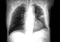
PA chest radiograph performed 5 months after mantle
radiation therapy of the mediastinum for treatment of Hodgkin's disease, shows typical
consolidation and air bronchograms strictly confined to the areas corresponding to the
radiation field. |
|
CT scan performed 8 weeks after mantle radiation
therapy for treatment of Hodgkin's disease shows ground-glass opacification confined to
the field of irradiation. |
Background: Radiation pneumonitis is an
interstitial pulmonary inflammation that can develop
in as many as 5-15% of patients with
thoracic irradiation, most often due to lung cancer, breast cancer, lymphoma, or thymoma.
Acute radiation pneumonitis occurs within 1-6 months following treatment. Symptoms can
include low-grade fever, cough, and fullness in the chest. Severe reactions can result in dyspnea, pleuritic chest pain, hemoptysis, acute respiratory distress, and death. Fibrosis
can occur without previous pneumonitis but once pneumonitis occurs, fibrosis is almost
certain to take place. The radiographic hallmark of radiation pneumonitis is a diffuse
infiltrate corresponding to a previous radiation treatment field.
Pathophysiology: Two separate and distinct mechanisms are
involved in the pathogenesis of acute radiation pneumonitis.The first, classical radiation
pneumonitis, involves direct toxic injury to endothelial and epithelial cells from the
radiation, resulting initially in an acute alveolitis. This process leads to an
accumulation of inflammatory and immune effector cells within the alveolar walls and
spaces. The accumulation of leukocytes distorts the normal alveolar structures and results
in the release of lymphokines and monokines. The alveolar macrophage is thought to play a
central role in the subsequent development of chronic inflammation (fibrosis). The second
mechanism, sporadic radiation pneumonitis, results in an "out-of-field"
response. This is thought to be an immunologically mediated process resulting in bilateral
lymphocytic alveolitis.
In contrast to acute radiation pneumonitis, permanent changes of radiation fibrosis can
take months to years to evolve but normally stabilize within 1-2 years. Pulmonary fibrosis
is the repair process that follows the acute inflammatory response and is characterized by
progressive fibrosis of the alveolar septa thickened by bundles of elastic fibers. The
process is not fully understood but believed to be a function of activation on cells to
produce cytokines and growth factors, which orchestrate most aspects of the inflammatory
response.
Frequency:
* In the US: Asymptomatic radiologic findings are observed in as many as 50% of treated
patients. Clinical radiation pneumonitis can develop in 5-15% of patients undergoing
radiation treatment to the thorax. The clinical pathologic course is biphasic and is
dependent upon the dose and volume of lung exposed and the use of chemotherapy agents.
Mortality/Morbidity: Morbidity and mortality vary greatly based on the volume of lung
irradiated, dose per fraction of radiation delivered, use of concomitant chemotherapy,
total dose of radiation delivered, and performance status of the patient. Predisposing
factors such as smoking history, collagen vascular disease, and steroid withdrawal also
affect the frequency of symptoms. Moderate-to-severe radiation pneumonitis occurs in an
estimated 2-9% of patients treated for lung cancer with combination chemotherapy and
irradiation. This represents the high-risk group. Even in this high-risk group, mortality
is estimated to be 1-2%.Race: No race predilection exists. Sex: Women tend to have higher
rates of moderate-to-severe radiation pneumonitis. This may reflect that most women have
smaller lung volumes and smaller forced expiratory volume in 1 second (FEV1) values. Thus,
given similar radiation field sizes, a greater proportion of lung may be at risk. This
also may represent an autoimmune predisposition to injury. Many autoimmune diseases, such
as systemic lupus erythematosus, are more common in women than in men and are a known risk
factor for increasing the chance of subsequent radiation-induced lung damage. Age: No
direct link to age exists. However, rates do increase as performance status decreases,
which is indirectly related to age.
Clinical Details: Classic radiation pneumonitis has 3 main
phases.
Early phase (first month): This represents a latent period of pneumonitis. During this
phase, loss of both type I and type II pneumonocytes occurs. Type II pneumonocytes produce
surfactant, and decreased amounts result in transudation of serum proteins into the
alveoli. This leads to edema of the intersitial spaces.
Intermediate phase (1-6 months): This is characterized by dose-dependent leakage of
proteins into the alveolar space, thickening of the alveolar septa, and development of
clinical symptoms. Common clinical symptoms include nonproductive cough, low-grade fever,
tachycardia, and dyspnea.
Late phase (6 months and later): This is characterized by a loss of capillaries and
increased collagen deposition. This results in restrictive changes within the lung
characterized by reductions in vital capacity, lung volumes, diffusing capacity of lung
for carbon monoxide (DLCO), and total lung capacity. |
Chest Xray Findings: Chest radiographic
findings vary from normal or subtle hazy ground glass density to marked patchy or
homogenous consolidation. Air bronchograms are commonly present and volume loss of the
affected portion of the lung may be observed. There is usually a sharp boundary crossing
the normal anatomic structures without segmental or lobar distribution. Rarely, an entire
lung or both lungs are involved (adult respiratory distress syndrome [ARDS]).False
Positives/Negatives: False positives include recurrent disease, infection/pneumonia,
cardiac disease, and lymphangitic carcinomatosis.
CT Scan Findings: Acute radiation pneumonitis changes,
especially the subtle ground glass opacity (GGO), are seen earlier on CT than on
radiographs. Different patterns of radiation-induced damage observed on chest radiographs
are seen to better advantage with CT and vary from early homogenous, slight increase in
radiodensity (GGO), to patchy or homogenous consolidation. Small pleural and pericardial
effusions are not uncommon.CT is useful in detecting recurrent tumor in an irradiated
area. This is suggested by the presence of a masslike lesion or development of focal air
space opacity without air bronchogram.CT scans can be used to detect and calculate volumes
of lung affected as a percentage of the total lung volume.
Intervention: Corticosteroids remain the treatment of
choice for radiation pneumonitis.
Prophylactically administered corticosteroids have been shown to decrease the physiologic
effects of radiation in mice. However, in human studies, this approach has failed to
prevent the development of clinical pneumonitis. TGF-B has been the target of more recent
studies. Angiotensin-converting enzyme (ACE) inhibitors have been shown to decrease the
expression of TGF-B1 in animals and recently have been tested in human trials. Researchers
at Duke University recently reviewed the records of 213 patients receiving thoracic
irradiation for lung cancer with curative intent. Of these patients, 12% were on ACE
inhibitors for hypertension. Initial results revealed that at the dose used for the
treatment of hypertension, ACE inhibitors had no protective effect. Future trials will
evaluate larger doses.The recommended treatment is to begin prednisone at 1 mg/kg as soon
as the diagnosis is reasonably certain. The initial dose is maintained for several weeks
and then reduced slowly. If steroids are tapered too soon or too quickly, exacerbation of
symptoms has been reported, requiring higher doses and longer treatment with steroids.
Antibiotics and anticoagulants have been evaluated as treatment options but neither has
been found to be clinically beneficial. Pentoxifylline has been shown to decrease late
effects from radiation damage but clinical trials in humans have shown no benefit. |