|
||||
|
Probability of late
rectal morbidity in 125I prostate brachytherapy |
|||||||||||||||||||||||||
|
|||||||||||||||||||||||||
|
|
||||
|
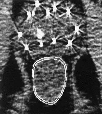
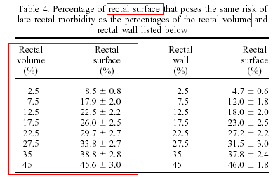
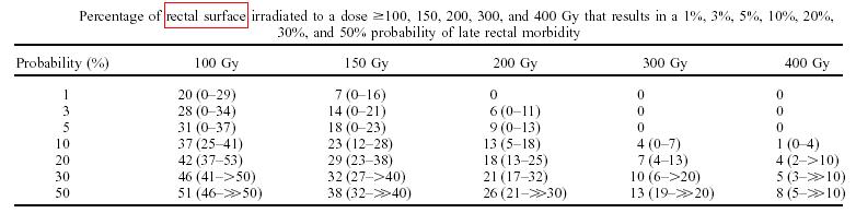
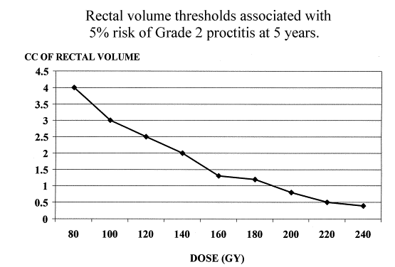
Purpose: Rectal toxicity is a concern in prostate brachytherapy because it is difficult to avoid delivering a dose equal to, or greater than, the prescription dose to the anterior surface of the rectum. The purpose of this study was to define the probability that a patient will experience Grade 2 (bleeding/ulceration) late rectal morbidity after 125I prostate brachytherapy according to the rectal dosimetry.Ninety-eight consecutive patients who received monotherapy 125I prostate implants for treatment of Stage T1-T2, favorable-risk adenocarcinoma of the prostate were evaluated for Radiation Therapy Oncology Group Grade 2 late rectal morbidity. The rectal dosimetry was based on a CT scan obtained at 3–9 weeks after implantation. The rectum was contoured on each CT image between the base and apex of the prostate. A dose-surface histogram was compiled for each implant, and the relative surface area that received a dose was recorded. Merrick reported an analysis of the rectal dosimetry of 45 patients after prostate brachytherapy. They defined the rectal dose as the dose received by the anterior rectal mucosa, which was identified using a rectal obturator. The only rectal complication observed in these patients was mild, self-limited proctitis; hence, a dose response for late rectal morbidity could not be established. Instead, the authors focused on defining the dosimetric parameters that should preclude the incidence of late rectal morbidity. They concluded that as long as the average dose to the anterior rectal mucosa is maintained at about 85% of the prescribed dose, the maximum kept at <125% of the minimal peripheral dose, and the length of the rectum receiving 100% and 120% of the prescribed dose kept to less than approximately 10 mm and 5 mm, respectively, the incidence of serious rectal complications will be rare. Snyder reported that the risk of developing Grade 2 radiation proctitis (defined as rectal bleeding occurring at least once a week for a minimum period of 1 month) is a function of the volume of the rectal wall that receives a specific dose. Thus, the risk can be assessed from a dose-volume histogram of the rectal wall (DWH). The volume threshold reported for 5% risk of Grade 2 proctitis at 5 years was 3.0 cm3 at 100 Gy, 2.0 cm3 at 140 Gy, 1.3 cm3 at 160 Gy, 1.2 cm3 at 180 Gy, 0.8 cm3 at 200 Gy, 0.5 cm3 at 220 Gy, and 0.4 cm3 at 240 Gy. One limitation of this work is that one can only determine whether the risk is >5% or <5%. All patients who received monotherapy 125I prostate implants for treatment of T1-T2 prostate cancer during 1997, 1998, and 1999 and had a CT scan at least 3 weeks postoperatively were entered into the study. A minimum of 3 weeks was required because the rectal dose increases significantly as the postimplant edema resolves (14). Of the 111 implants performed during this period, 98 met this criterion. All patients had at least 15 months follow-up after implantation. The median follow-up was 32 months (range 15–54). Patients were evaluated for late rectal toxicity based on the Radiation Therapy Oncology Group/European Organization for Research and Treatment of Cancer late radiation morbidity scoring schema. All reported incidences of late morbidity were retrospectively confirmed by colonoscopy.The implants were preplanned to deliver a minimal dose of 150 Gy to the prostate plus a symmetric 3–5-mm dose margin (including the posterior margin). The seeds were peripherally loaded using 0.4–0.6-mCi 125I seeds. This peripheral loading produced an isodose distribution characterized by a broad dose minimum in the central portion of the prostate encircled by a high-dose region. The preplans were based on a preimplant CT scan, and the implants were performed under fluoroscopy guidance (15). The rectal postimplant dosimetry was calculated from a CT scan obtained 4–6 weeks after implantation using software developed in-house. For this purpose, the rectum was defined as the portion of the rectum between the base and the apex of the prostate (i.e., the rectum was only contoured on CT images that contained prostate). Results: Of the 98 patients, 10 developed Grade 2 late rectal morbidity. The percentage of the rectal surface that received 100, 150, 200, and 300 Gy was significantly greater (p ?0.02) for patients who experienced late rectal morbidity. The probability of late rectal morbidity increased with both the dose and the percentage of the rectal surface that received that dose. The probability was ?1% when 20%, 7%, and 0% of the rectal surface received 100, 150, and 200 Gy, respectively. The probability increased to ?5% when 31%, 19%, and 9% of the rectal surface received these doses. The probability of late rectal morbidity can also be expressed in terms of the maximal rectal dose. The probability of late morbidity was 0.4%, 1.2%, and 4.7% when the maximal rectal dose was 150, 200, and 300 Gy, respectively. Conclusion: The percentage of the rectal surface that receives a dose 100 Gy is predictive of Grade 2 (bleeding/ulceration) late rectal morbidity after 125I prostate brachytherapy. The probability of late morbidity depends on both the dose and the percentage of the rectal surface that received that dose. Our results indicate that the rectum can tolerate doses of 100, 150, and 200 Gy to approximately 30%, 20%, and 10% of the rectal surface with a ?5% risk of late morbidity. Our results also indicate that the practical guideline for limiting the incidence of late morbidity to 1%, 3%, or 5% is to keep the maximal rectal dose to <200, 250, and 300 Gy, respectively.
Defining the risk of developing Grade 2 proctitis following 125I
prostate brachytherapy using a rectal dose–volume histogram analysis
|
![]()