|
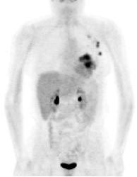
PET exam for the evaluation of locoregional recurrence: The patient shown in the case below had a history of breast cancer and had developed left chest pain. She presented for the evaluation of possible metastatic disease. The CT scan revealed extensive soft tissue thickening in the left breast which was felt possibly related to scar from prior surgery and radiation therapy. There was a 2 cm lymph node in the left axilla (not shown) which was concerning for metastatic disease. Axial (center) and coronal (right) images from the patients FDG PET exam demonstrated marked increased FDG accumulation within the left breast corresponding to the soft tissue abnormality on CT. There were also multiple foci of increased uptake within the the left axilla. Biopsy revealed recurrent breast cancer. |
|
 |

