A population-based atlas and clinical target volume for the head-and-neck lymph nodes 
Ian Poon IJROBP Volume 59, Issue 5, Pages 1301-1311 (1 August 2004)

neck_nodes_ucsf.jpg (9764 bytes)

(a–l) Marked lymph nodes (LNs) with each LN group separated by color.


Light blue, submental LN (Ia);
dark blue, submandibular LN (Ib);
yellow, jugular LN (II, III, IV);
red, spinal accessory LN (V);
light green, retropharyngeal LN;
orange, parotid LN;
dark green, anterior jugular LN (VI);
pink, retroauricular/occiptal LN.

click on letter to see pics
Level           Distinguishing features                                                                            Superior limit               Inferior limit

A
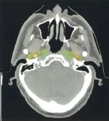
                  Nasopharynx                                                                                       Sphenoid sinus             Hard palate
                     
B            Maxillary alveolus/hard palate                                                                    Hard palate               Foramen magnum
                      
C    Upper oropharynx/ascending ramus of mandible; parotid gland                       Foramen magnum    C2 vertebrae
                    
D, E    Mid/lower oropharynx, submandibular gland, inferior to parotid gland         C2 vertebrae    Vallecula
                    
F    Thyroid cartilage, glottic structures                                                              Thyroid cartilage    Cricoid cartilage
                   
G    Thyroid gland                                                                           Superior limit of thyroid gland    Inferior limit of thyroid gland       
           
H    Apices of lungs, clavicles                                                                  Apex of lung    Head of clavicle
                   
Jaw 1    Ascending ramus of mandible , Jaw 2     50% of bony mandible , Jaw 3    <25% of mandible , Jaw 4     Inferior to mandible