|
Multiple Myeloma / Plasmacytoma
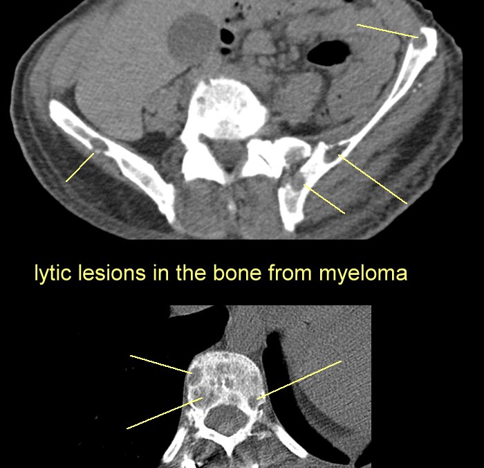
Images of myeloma in the bone: (note that the goal is to vigorously treat the myeloma so
that the bones do not become this brittle.)
#1
Radiographs Showing Multiple, Irregular
Lytic Lesions in the Skull (Panel A) and Pelvic Bones (Panel B). Similar lesions were
present in the ribs, axial skeleton, and extremities
#2 Large Destructive Lesion in the
Right Iliac Bone
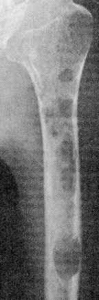
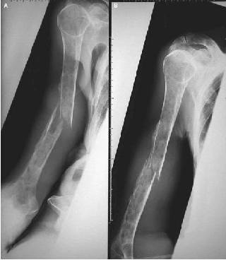
#3 Pathologic Fracture of the Right
Humerus
#4 Pathologic fracture of the
humerus
#5 Diffuse lytic
bone lesions in pelvis and femurs
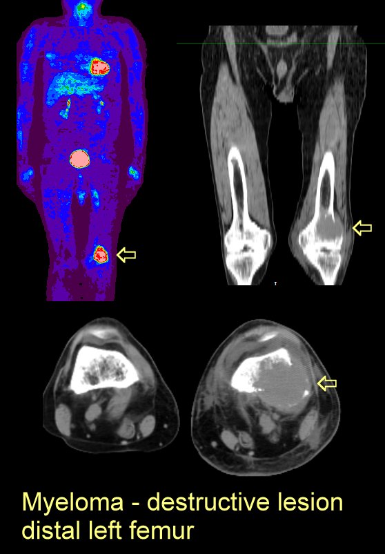
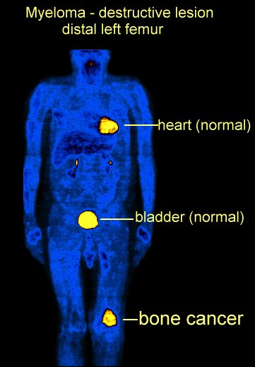
#6 Large destructive lesion in distal left femur on PET
and CT: here and
here
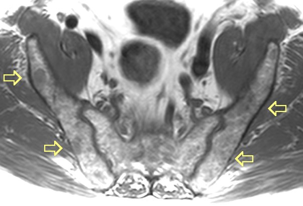
#7 MRI images of very diffuse disease
here and
here
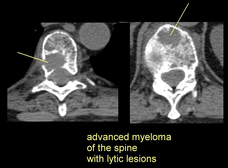
#8 Advanced disease in the thoracic spine
here and
here
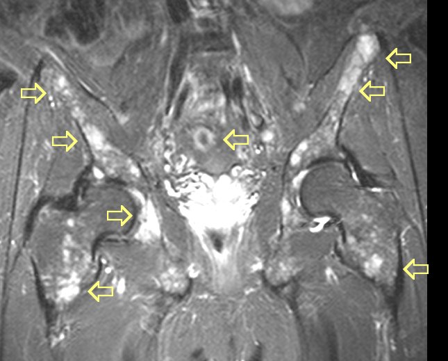
#9 Advanced disease in the right hip and pelvis:
here,
here, and here
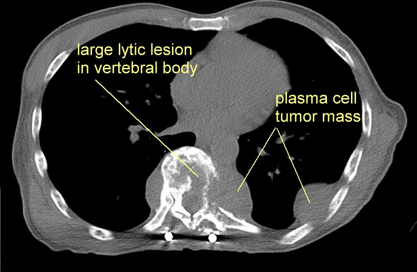
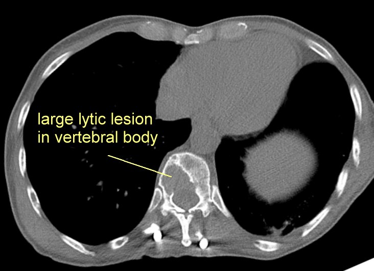
#10. Myeloma can make small punched out holes in the bone
(called lytic lesions) or form large masses (plasma cell tumors) go
here, here and
here
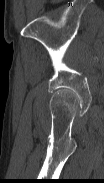
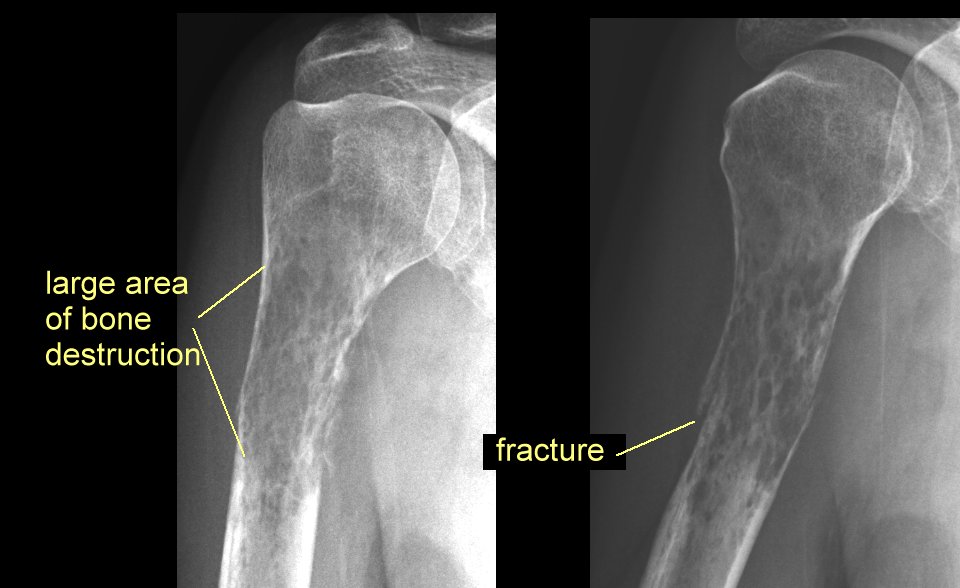
#11. Myeloma may destroy so much bone that a fracture
results, go here
#12 Punched out skull lesions are classic for myeloma, go
here
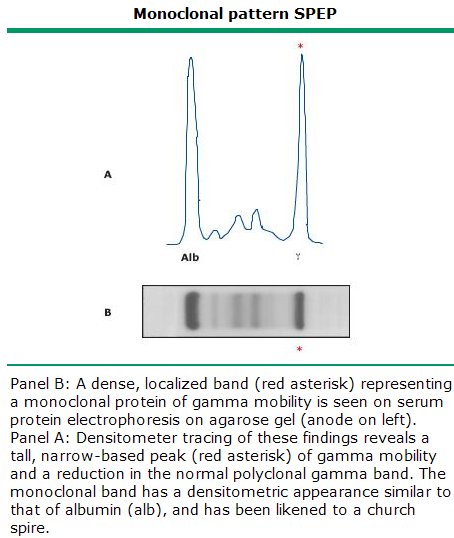
other images: the blood smear may be a tip off
of myeloma (go here) the most common test
is the serum protein electrophoresis (SPEP) go
here and the immunofix test (here)
with the final diagnosis a bone marrow biopsy (here) |
