 |
Review - Merkel Cell Carcinoma (MCC)
|
|
Review - Merkel Cell Carcinoma (MCC)
|
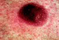
IN 1875, FRIEDRICH Sigmund Merkel (1845-1919) described a unique epidermal nondendritic, nonkeratinocyte cell, which he called a tactile cell (Tastzelle).Merkel cells are now generally believed to be primary neural cells, found as single cells within the basal layer of the epidermis or grouped together as a component of the tactile hair disc of Pinkus in the hair-bearing skin of mammals, functioning as slowly adapting type I mechanoreceptors. MCC is a rare cutaneous neoplasm of the elderly population with estimated 470 new cases in the United States each year; this incidence figure compares with 31,000 new cases of melanoma annually. Although most studies show a male predominance, with SEER data suggesting a ratio of 2.3:1, other case series demonstrate a slightly higher incidence in women.MCC is a disease of the elderly with an average age at the time of diagnosis of 69 years; only 5% of all reported patients have been diagnosed below the age of 50 years. Clinical Presentation Most patients with MCC present with localized disease (70% to 80%); 10% to 30% of patients have regional lymph node involvement, and 1% to 4% have distant metastases at the time of initial presentation There is no accepted staging system for MCC, but several investigators have adopted a simple system proposed by Yiengpruksawan et al:stage I, localized skin disease (stage IA 2 cm, IB > 2 cm); stage II, regional lymph node disease; and stage III, metastatic disease. Purported favorable prognostic factors include primary tumor location in the head and neck region and without involvement of regional lymph nodes, tumors 2 cm in size (stage IA), and female sex. Initially, MCC was thought to be a cancer with a good prognosis, as only one out of the first five patients described by Tokersuccumbed to the disease, and only three deaths occurred in the first 24 reported cases. However, MCC has subsequently been shown to be a highly aggressive and lethal tumor, comparable with small-cell lung cancer and melanoma in its behavior with regards to recurrence, metastatic spread, and mortality. The overall recurrence rate ranges from 55% to 79%, occurring most often locally or in regional lymph nodes, with the majority of recurrences appearing within the first 6 to 12 months after initial diagnosis. |
| Stage | Median Survival | Survival/5y | Treatment | |
| I | localized | na | 64% | local excision with >2cm margin, sentinel node, postOp XRT (45-50Gy) |
| IA | 2cm | 30 mos | ||
| IB | > 2cm | 26 mos | ||
| II | nodes involved | 18 mos | 47% | surgery with node dissection, postOp XRT to tumor bed and nodes |
| III | metastases | 5 mos | 0% | palliative XRT + chemo (CAV or EP) |
![]()