|
|
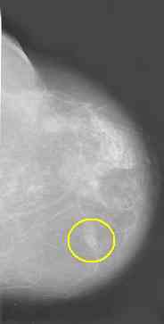
Basic Information About Mammography in younger women with more density in their breasts, MRI may be more sensitive than mammograms (see studies , here and here) It's important to emphasize that mammograms may miss early breast cancers. The sensitivity in younger women is particularly poor. The NCI states that: "Overall sensitivity is approximately 75% but ranges from 54% to 58% in women younger than 40 years to 81% to 94% in those older than 65 years."
mammogram showing
cancer#1 and #2
see large image database
here see
NCI
on mammograms ,
ACS
info Upper picture is mammograms of younger woman (Bilateral mediolateral oblique views in a 45-year-old woman. These breasts are a mixture of dense fibroglandular tissue (white) and radiolucent fat (dark gray). The pectoral muscles (arrows) are seen at the posterior aspect of the breast. A small carcinoma could easily be obscured within one of the dense tissue areas.) Middle picture is mammogram of older woman (Bilateral mediolateral oblique views of a 65-year-old woman. Almost all fibroglandular tissue has been replaced by radiolucent fat. A small tumor would be visible at any location in this breast, unless it were not positioned properly over the film} Lower Picture show appearance of Cancer (Ductal carcinoma manifested by a spiculated mass (*) and clusters of fine, malignant calcifications (arrows) both within and outside the tumor. The mass was an invasive carcinoma, with the calcifications representing extensive intraductal extension.) There is variation among normal women as to breast density. The more dense the breast the higher the risk of cancer and the more difficulty seeing cancer on mammograms. The radiographic appearance of the female breast varies among women of the same age because of differences in tissue composition. Fat is radiographically lucent and appears dark on a mammogram, whereas connective and epithelial tissues are radiographically dense and appear light — an appearance that we refer to as "mammographic density." Examples are shown in Figure 1. Wolfe first described an association between a qualitative classification of dense mammographic patterns and an increased risk of breast cancer. At least 15 other cohort studies have confirmed this association. Ten studies (six case–control studies and four cohort studies involving a total of 4747 cases of breast cancer have assessed mammographic density quantitatively. All found a risk of breast cancer in the category with the most extensive dense tissue that was 1.8 to 6 times as high as that in the category with the least extensive dense tissue, and in eight of these studies, the risk was at least quadrupled. |
