Curative radiotherapy for
primary orbital lymphoma
Bhatia S, Paulino AC, Buatti JM, Mayr NA, Wen B C International Journal of Radiation
Oncology*Biology*Physics
01 November 2002 (Vol. 54, Issue 3, Pages 818-823)Orbital lymphoma is rare and accounts for <1% of all cases of non-Hodgkin’s
lymphoma. Most institutional reports had limited numbers of patients . Various subsites within the orbit may be involved, including the
conjunctiva, lacrimal apparatus, eyelids, and musculature. Early-stage orbital lymphoma is
best treated with radiotherapy (RT). In a study by Esik, the 10-year local
relapse-free survival and 20-year cause-specific survival rates were 100%, 0%, and 42% and
100%, 67%, and 0%, respectively, for patients primarily treated with RT, surgery, or
chemotherapy. The purpose of the present report was to review the efficacy of RT on
early-stage primary orbital lymphoma as given in our institution and to determine the late
effects and complications after treatment.
Treatment characteristics
All patients were treated with curative intent using external beam RT alone after biopsy
of the lesion. Treatments were given 5 d/wk using a fraction size of 200 cGy in 36 orbits,
180 cGy in 15, and 170 cGy in 1. For low-grade lymphomas, the
median dose was 3000 cGy (range 2000–4020); for the intermediate- and high-grade
tumors, the median dose was 4000 cGy (range 3000–5100). Beam energy was
4-mV photons in 34 (65%), 6 mV in 6 (12%), 1.25 mV in 4 (8%), 10 mV in 3 (6%), and
orthovoltage RT in 2 (4%). Electron beam RT was used in 3 patients (6%). The 2 patients
who received kilovoltage RT were treated during the earlier part of the study. A
lens-sparing approach was used in 19 (37%) of the orbits treated. Six patients were
treated with parallel-opposed lateral fields; five had bilateral lymphoma and one had
lymphoma and contralateral atypical hyperplasia of the orbit. The lenses of the 6 patients
were spared from RT by using a half-beam block technique centered at the outer fleshy
canthus of the eyes. The other eight orbits treated with a lens-sparing approach either
had a lead contact lens shield (7 cases) or a hanging lens block (1 case) in the AP field.
Methods and Materials: Between 1973 and 1998, 47 patients (29 women [62%] and 18 men
[38%], median age 69 years, range 32–89) with Stage IAE orbital lymphoma were treated
with curative intent at one department. Five had bilateral orbital involvement. The tumor
was located in the eyelid and extraocular muscles in 23 (44%), conjunctiva in 17 (33%),
and lacrimal apparatus in 12 (23%). The histologic features according to the World Heath
Organization classification of lymphoid neoplasms was follicular lymphoma in 25,
extranodal marginal zone B-cell lymphoma of mucosa-associated lymphoid tissue type in 8,
diffuse large B-cell lymphoma in 12, mantle cell lymphoma in 6, and peripheral T-cell
lymphoma in 1. For the purposes of comparison with the existing literature on orbital
lymphomas, the grading system according to the Working Formulation was also recorded. The
histologic grade was low in 33 (63%), intermediate in 18 (35%), and high in 1 (2%). All
patients were treated with primary radiotherapy alone. The median dose for low-grade
tumors was 3000 cGy (range 2000–4020); the median dose for intermediate and
high-grade tumors was 4000 cGy (range 3000–5100). A lens-sparing approach was used in
19 patients (37%). Late complications for the lens and cornea were scored according to the
subjective, objective, management, and analytic (SOMA) scale of the Late Effects of Normal
Tissue (LENT) scoring system. The median follow-up was 55 months (range 6–232).
Results: The local control rate was 100% in the 52 orbits treated.
The 5-year overall survival and relapse-free survival rate was 73.6% and 65.5%,
respectively. Tumor grade and location did not predict for overall survival or
relapse-free survival. Seven patients (15%) developed distant recurrence (brain 2,
extremity 2, mediastinum 1, liver 1, and retroperitoneum 1). One patient (2%) developed
cervical node metastasis. The 5- and 10-year cataract-free survival rate was 56.7% and
32.9%, respectively. Of the 12 lens complications, 8 were LENT Grade 1 and 4 were Grade 3
toxicity. Only male gender predicted for an increased risk of cataract formation.
Radiotherapy dose and technique did not predict for cataract formation; however, none of
the patients who underwent the lens-sparing technique developed Grade 3 lens toxicity or
required surgical correction. Of the nine corneal events, two were Grade 1, four Grade 2,
and three were Grade 3 toxicity. Ten dry eyes were recorded; all were mild, and no patient
had severe dry eye syndrome. Neovascular glaucoma was seen in 1 patient. No injury to the
retina or optic nerve was reported.
Discussion
Orbital lymphoma is a rare type of malignancy. As
a result, most of the current literature consists of single-institution retrospective
reviews. In this report, we present one of the largest single-institution experiences in
early-stage orbital lymphoma treated with curative intent. Other studies have had a
smaller number of cases and some have included more advanced or palliative cases.
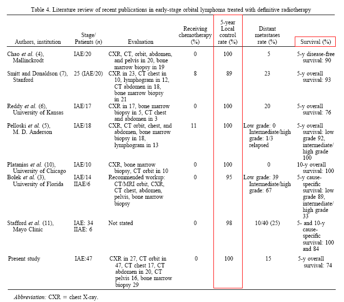
Local control with RT for orbital lymphoma is excellent. Table 4 summarizes the modern
literature concerning orbital lymphomas. Local control rates have
ranged from 89% to 100%, with a distant metastases rate of 0–25%. Our
experience showed that 15% of patients developed distant relapse, consistent with
previously reported studies. The only exception was a study from the University of Florida
that showed a 39% and 67% distant metastasis rate for low- and intermediate/high-grade
tumors, respectively.
We did not find a statistically significant
difference in relapse-free survival among low-grade and intermediate/high-grade tumors;
however, the median relapse-free survival time was longer in those with low-grade tumors
(13.92 vs. 7.17 years). A larger number of patients may have shown a statistical
difference.
The staging workup for orbital lymphoma is controversial. What tests should be required to
evaluate a patient with orbital lymphoma? Table 4 shows the different tests used at
various institutions. Most of the studies had inconsistent staging tests, with the
exception of two . The M. D. Anderson study used chest X-ray, CT scan of the orbit, CT
scan of the chest and abdomen, and bone marrow biopsy in all 18 patients. The 5-year local
control rate was 100%, with a 0% distant metastasis rate for low-grade tumors. One of 3
patients with intermediate/high-grade tumor developed a distant relapse. The Mallinckrodt
study included chest X-ray and CT of the orbit and of the abdomen and pelvis in all
patients and a bone marrow biopsy in all but 1 patient. The local control rate was 100%,
with a 5% distant metastasis rate. Could the excellent results in the M. D. Anderson and
Mallinckrodt institutes be a function of patient selection, with only the true Stage I
patients analyzed? The patterns of failure in our study do not support this hypothesis. Of
the eight distant and regional metastasis sites, five (brain 2, extremity 2, cervical node
1) would not have been found by CT of the chest, abdomen, or pelvis or bone marrow biopsy.
The median time to relapse was 36 months (range 6–149), suggesting that most, if not
all, of these relapses were not present at the initial diagnosis. Furthermore, excellent
results have been reported by other studies with “incomplete” staging).
Exactly what dose should be used for low-grade and intermediate-grade tumors is not clear
in the present study, because all cases were locally controlled. Follicular lymphomas and
extranodal marginal zone B-cell lymphomas of the MALT type were treated with a median dose
of 3000 cGy (range 2000–4020), and diffuse large B-cell lymphomas and mantle cell
lymphomas had a median dose of 4000 cGy (range 3000–5100). We
recommend 3000 cGy for follicular and extranodal marginal zone B-cell lymphomas of the
MALT type and 3600 cGy in conventional fractionation for diffuse large B-cell and mantle
cell lymphomas. It is possible that doses <3000 and 3600 cGy may be enough.
Four follicular or extranodal marginal zone B-cell lymphomas received doses of 2000, 2000,
2880, and 2975 cGy, given in conventional fractionation, and were locally controlled. Nine
of 19 diffuse large B-cell or mantle cell lymphomas received 3000–3960 cGy (3000 cGy
in 4, 3600 cGy in 4, and 3960 cGy in 1), and 10 received doses of 4000–5100 cGy in
conventional fractionation and were locally controlled.
Bolek treated 12 patients with low-grade lymphoma
that were locally controlled with doses ranging from 1500 to 2600 cGy using
156–200-cGy fractions. Smitt and Donaldson only had 1 patient with low-grade
lymphoma receiving 2800 cGy; this patient subsequently developed a relapse and was
salvaged by surgical excision. At the M. D. Anderson Cancer Center, 1 case of
intermediate-grade lymphoma received 3600 cGy in 18 fractions without chemotherapy and was
controlled; one orbit with diffuse large cell lymphoma received 3060 cGy in 17 fractions
with chemotherapy and was controlled.
Most complications in our experience were mild, requiring no
intervention. No cases of retinopathy, optic nerve injury, or severe dry eye syndrome were
reported. Men were found to be more likely to develop cataracts after RT than were women.
The RT dose, fractionation, beam energy, beam arrangement, lens-sparing technique, and use
of concurrent steroids were not statistically related to cataractogenesis. Cataracts were
found in 12 lenses, two-thirds of which were not symptomatic and were found only on
routine follow-up. Four lenses, however, required surgical intervention. All four Grade 3
lens toxicities developed in patients who had been treated without a lens-sparing
technique. Because of the higher grade of cataracts in those treated
without a lens-sparing technique, we recommend a lens-sparing approach whenever feasible.
The available literature is presented in Table 5 and supports this approach. Of 76 and 103
cases treated with or without lens shielding, 13% and 27%, respectively, developed a
cataract (p = 0.023)
Three patients had Grade 3 corneal toxicity, all
with ulceration and requiring topical antibiotics. Because the primary function of the
cornea is refraction, an alteration in corneal shape such as ulceration and scar formation
can cause significant refractive changes and alter vision. However, none of the three
required surgical intervention. The tolerance dose of the cornea
has been reported to be 5000 cGy in conventional fractionation, although some
have indicated that a dose of 3000 cGy can result in keratitis if larger than conventional
fractionation is used. All our patients who developed corneal ulceration received <5000
cGy with conventional fractionation.
One patient developed neovascular glaucoma. This is an uncommon RT complication caused by
abnormal vascularization and subsequent closure of normal aqueous outflow channels
resulting in increased intraocular pressure. Neovascular glaucoma is more commonly seen in
patients treated with plaque RT). Takeda and colleagues have previously reported a
14% incidence of neovascular glaucoma in those receiving >5000 cGy external beam RT to
the iris.
The issue of bilaterality in orbital lymphoma is interesting. In our study, 5 (11%) of 47
patients had bilateral disease. At Stanford University, researchers have found a 25%
incidence of bilaterality, and at University of Florida, 10% of localized orbital lymphoma
developed a metachronous, contralateral orbital lymphoma at 3.8 and 5.5 years . Reddy
found a 24% incidence of bilateral disease in 17 patients. Others have not reported
any bilateral cases
Conclusion: Radiotherapy alone is a highly effective modality in the curative management
of primary orbital lymphoma. Most complications were minimal and did not require medical
or surgical intervention. Although the use of the lens-sparing technique did not influence
the incidence of cataractogenesis, we continue to recommend this approach whenever
possible, because our experience indicates a higher grade of toxicity occurs and a higher
incidence of corrective surgery is needed in patients treated without lens protection.
|