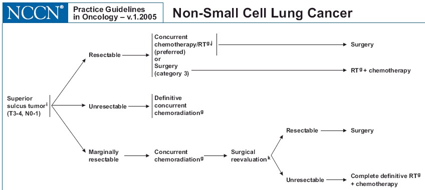
| Some lung tumors invade into the chest wall or ribs and are considered marginally resectable. The NCCN guidelines recommend preoperative radiation or chemoradiation followed by surgery (see NCCN.) A mores specific tumor in the upper part of the chest cavity that impinges on nerves is called a Pancoast tumor and is generally treated with preoperative radiation +/- chemotherapy and then surgery as noted below: |
| Pancoast tumors cause symptoms
due to their anatomic location in the superior sulcus of the lung. The clinical syndrome
was first described by Edwin Hare, however, it was named after Henry Pancoast who defined
the findings in 1932. This clinical syndrome is due to tumor growth in the superior sulcus
at the lung apex and is related to invasion and destruction of neural structures in the
area icon gif. Pancoast's syndrome is commonly manifested by: * pain in the shoulder or medial portion of the scapula. * radicular pain with or without muscle wasting in the distribution of the ulnar nerve to the elbow (T1 distribution) and/or medial forearm and hand (C8 distribution). * Horner's syndrome (ptosis, miosis, hemianhydrosis, enopthalmus). Although this is a lung tumor, pulmonary symptoms are rare. The most common initial presentation is pain localized to the shoulder which is found in 90% of cases. Consequently, many cases are initially misdiagnosed as shoulder arthritis. Continued tumor growth leads to sympathetic chain involvement with a Horner's syndrome in 62% of cases. Because these tumors commonly invade the chest wall, they are staged as T3. Other common findings at presentation include invasion or destruction of ribs in 45% of cases and invasion or destruction of vertebrae in 23%. The most common cell type causing Pancoast syndrome is squamous cell carcinoma (52%) with adenocarcinoma and large cell (both 23%) being less common causes. Small cell carcinoma is a rare cause of Pancoast syndrome (1%). Radiographically the tumor is often difficult to see and may only be visible as a vague cap at the lung apex, similar to pleural thickening. CT and MRI are helpful in determining the extent of involvement and invasion of adjacent structures. MRI more clearly delineates tumor extension than CT primarily because MRI offers coronal and sagittal imaging with superior definition of vascular and neural structures. These more advanced imaging techniques are the key to determining resectability which requires accurate definition of vascular, neural, and bony invasion.Pancoast tumors are often difficult to diagnose histologically. Bronchoscopic biopsy only rarely provides diagnostic tissue samples due to the peripheral location of these tumors. In addition to the degree of local invasion, nodal staging is an important prognostic factor. Mediastinoscopy is useful to detect the presence of mediastinal metastases. Those patients staged N0 have a better prognosis than those with nodal metastases. Today, the standard of care for patients with a Pancoast tumor is radiation followed by tumor and chest wall resection when feasible. There are no definite criteria for unresectability, but the following general guidelines have been proposed: * N2 nodal disease * extensive vertebral body invasion * distant metastases * superior vena cava syndrome. Retrospective study has suggested that radiotherapy followed by surgical resection is the best treatment available for operable patients with T3 N0 or N1 disease. For unresectable patients radiotherapy is important for palliative care, but rarely produces a long term cure (6% survival at 5 years). |