 |
Preoperative Staging of Non-Small-Cell Lung Cancer with
Positron-Emission Tomography
Remge M. Pieterman, John W.G. van Putten, Jacobus J. Meuzelaar, Eduard L. Mooyaart,
Willem Vaalburg, Gerard H. Koeter, Vaclav Fidler, Jan Pruim, Harry J.M. Groen
We prospectively compared the ability of a standard approach to staging (computed
tomography [CT], ultrasonography, bone scanning, and, when indicated, needle biopsies) and
one involving PET to detect metastases in mediastinal lymph nodes and at distant sites in
102 patients with resectable non-small-cell lung cancer. The presence of mediastinal
metastatic disease was confirmed histopathologically. Distant metastases that were
detected by PET were further evaluated by standard imaging tests and biopsies. Patients
were followed postoperatively for six months by standard methods to detect occult
metastases. Logistic-regression analysis was used to evaluate the ability of PET and CT to
identify malignant mediastinal lymph nodes.
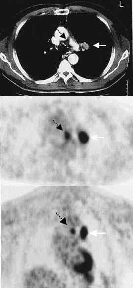
Results. The sensitivity and specificity of PET for
the detection of mediastinal metastases were 91 percent and 86 percent, respectively.
The corresponding values for CT were 75 percent and 66 percent. When the results of
PET and CT were adjusted for each other, only PET results were positively correlated with
the histopathological findings in mediastinal lymph nodes. Conclusions. PET
improves the rate of detection of local and distant metastases in patients with
non-small-cell lung cancer. (N Engl J Med 2000;343:254-61.) |