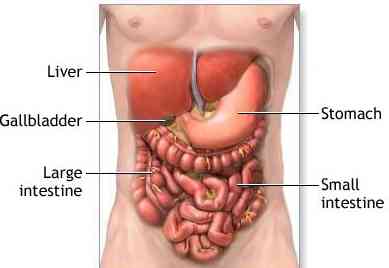
Liver Anatomy and
Metastases Images
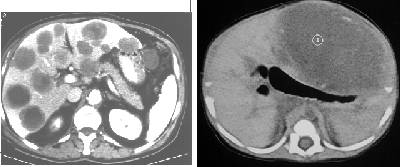
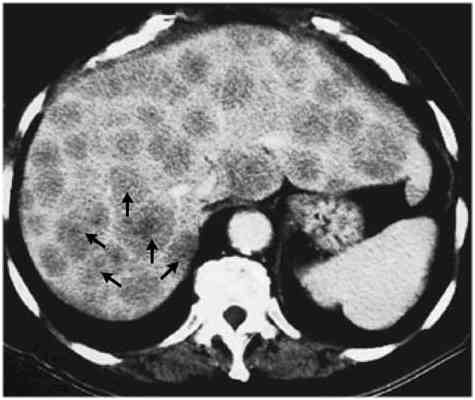
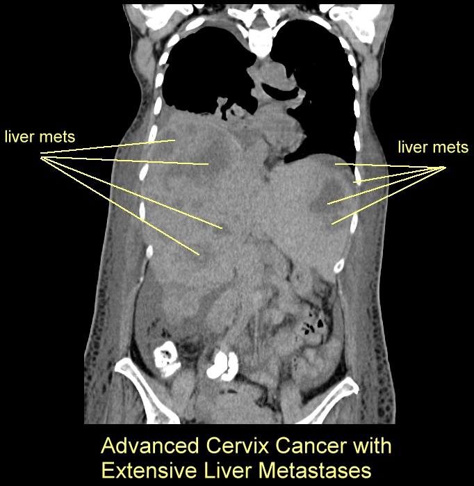
picture at left CT shows multiple liver metastases throughout the liver and below CT
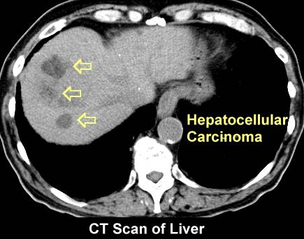
with large metastasis in the right lobe of the liver
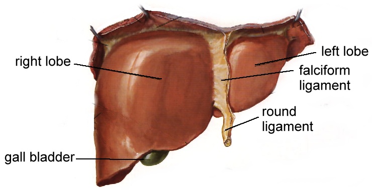
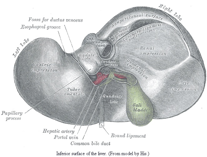
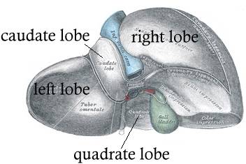
normal liver,
more anatomy,
more, superior
view, inferior view, posterior-inferior view,
segmental anatomy and
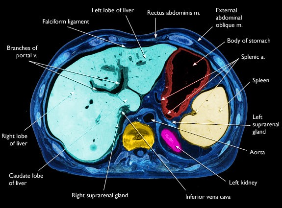
see CT of the normal liver,
CT normal liver, CT with multiple liver mets,
CT advanced liver mets
cross section through liver,
Primary liver cancers (hepatoma) can look like liver mets (go here).
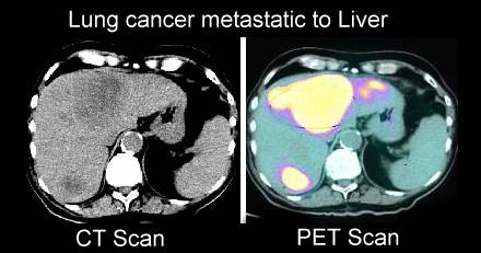
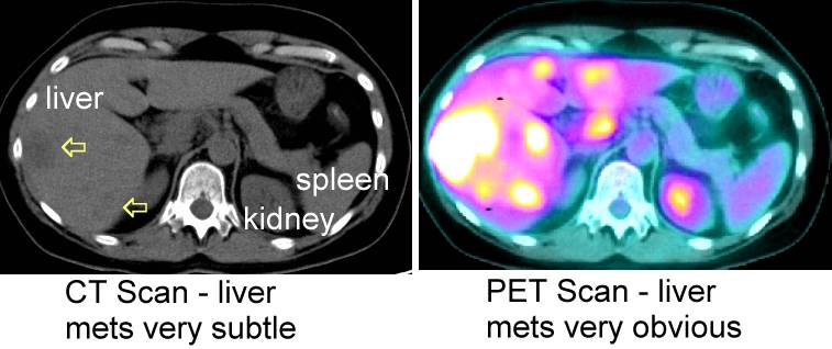
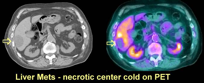
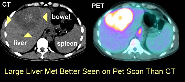
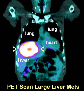
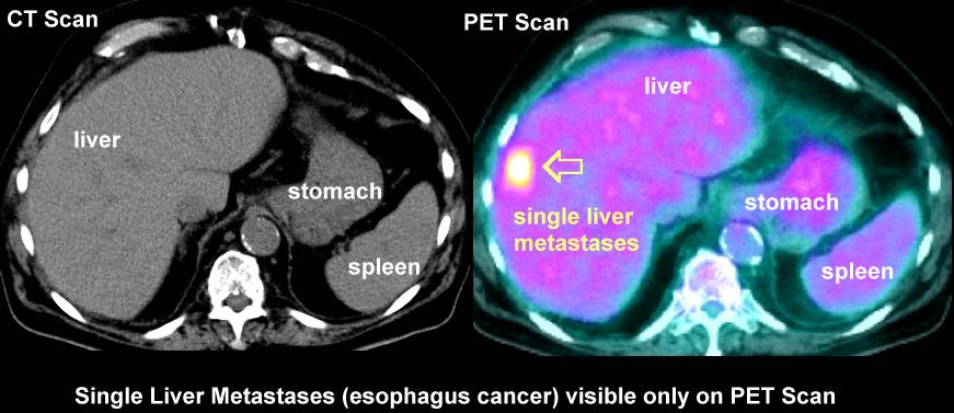
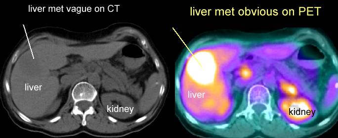
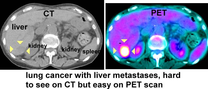
PET
scans are very good for liver mets (here, here, here, here , here
here, here,
here,
here,
here and here).
|
