Frequency:
Mortality/Morbidity: KA is believed to
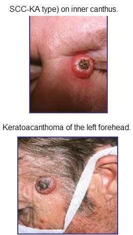
have a good prognosis; however, it recently was reclassified as SCC-KA
type to reflect the difficulty in histologic differentiation, as well as
the uncommon but potentially aggressive nature of KA. KA infrequently
presents as multiple tumors and may enlarge (5-15 cm), become aggressive
locally, or rarely, metastasize.
Race: KA is less common in
darker-skinned individuals.
Sex: Male-to-female ratio is 2:1.
Age: KA has been reported in all age
groups, but incidence increases with age. KA is rare in persons younger
than 20 years.
History: KA typically grows rapidly,
attaining 1-2 cm within weeks, followed by a slow involution period
lasting up to 1 year and leaving a residual scar if not excised
preemptively. Since expedient therapy almost always is instituted, the
true natural course of the tumor cannot be confirmed with
certainty.
Physical:
- Most KAs occur on sun-exposed areas. The face,
neck, and dorsum of the upper extremities are common sites. Truncal
lesions are rare.
- Lesions usually are skin-colored to pinkish-red.
Unaffected skin retains its normal appearance.
Causes:
- The definitive cause of KA remains unclear;
however, several potentiating factors should be considered.
- Epidemiologic data of KA is notably similar to SCC
and Bowen disease (BD; SCC in situ) concerning age, sex, and the
anatomic site of lesions. This data strongly supports a common etiology
among KA, SCC, and BD. Epidemiologic data support sunlight as an
important etiologic factor.
- Industrial workers exposed to pitch and tar have
been well established as having a higher incidence of KA, as well as
SCC.
- A recent study suggested a strong association
between cigarette smoking and the development of KA.
- Trauma, human papilloma virus (specifically types
9, 11, 13, 16, 18, 24, 25, 33, 37, and 57), genetic factors, and
immunocompromised status also have been implicated as etiologic
factors.
- Recent work has identified that up to one third of
keratoacanthomas harbor chromosomal aberrations. Recurrent aberrations
include gains on 8q, 1p, and 9q with deletions on 3p, 9p, 19p, and 19q.
One other report identified a 46,XY,t(2;8)(p13;p23) chromosomal
aberration.
Procedures:
Histologic Findings:
KAs are composed of singularly well-differentiated squamous
epithelium that show only a mild degree of pleomorphism and likely form
masses of keratin that constitute the central core of KA.
Pseudocarcinomatous
infiltration in KA typically presents a smooth, regular, well-demarcated
front that does not extend beyond the level of the sweat glands.
The term SCC-KA type has been
introduced for otherwise classic KAs that reveal a peripheral zone formed
by squamous cells with atypical mitotic figures, hyperchromatic nuclei,
and loss of polarity to some degree. These marginal cells also may
penetrate into surrounding tissue in a more aggressive
pattern.
Medical Care: Treatment of KA is
primarily surgical. Reserve medical treatment for exceptional cases where
surgical intervention is either not feasible or desirable. For example,
medical intervention may be appropriate in patients with multiple lesions,
in lesions not amenable to surgery because of size or location, and in
patients with comorbidities that dissuade surgical procedure.
Systemic retinoids, such as
isotretinoin, are a consideration for patients with lesions too numerous
for surgical intervention.
Intralesional methotrexate,
5-fluorouracil, bleomycin, and steroids have been used with success in
patients who are either poor surgical candidates or have lesions not
amenable to surgery because of size or location. Both topical imiquimod
and 5-fluorouracil have been used with anecdotal success.
Note much of the literature
concerning medical intervention for KA is limited to case reports and of
unproved efficacy. Be cautious when making the decision to pursue medical
in lieu of surgical intervention and perform appropriate
follow-up.
Surgical Care:
|