Epidemiologic studies indicate that these lesions are more common in
persons of Hispanic descent and in woman. Some neurosurgeons recommend that family members
of patients with cavernous malformations have a screening MR scan of the brain. Surgery
for these lesions is indicated only if they have bleed or are likely to bleed and if the
lesions are located in a surgically accessible area. Cavernous malformations in areas of
"eloquent brain" or in the brainstem are usually observed (and not operated)
unless they have bled at least twice.
The majority of patients with cerebral cavernous malformations present with headaches,
seizures, or neurologic deficits from hemorrhage. Many of these malformations are
identified in patients who undergo brain imaging for unrelated symptoms. Most patients
with incidentally discovered malformations or those with minimal symptoms can be followed
with periodic imaging studies unless new neurologic symptoms occur . Some malformations
that have caused hemorrhage or are associated with intractable seizures are suitable for
microsurgical resection. Occasional cavernous malformations are located in brain locations
associated with a high risk during resection. For such patients alternative therapies are
desirable. When malformations are located within the parenchyma of the brain stem or
diencephalon, stereotactic radiosurgery should be considered. For patients with lobar but
critically located malformations (e.g., those within motor cortex), for those that have
undergone partial resection, or for those patients who refuse surgery and have progressive
symptoms, radiosurgery may also be considered.
Reports of successful microsurgical extirpation of cavernous malformations were noted more
frequently in the literature after 1985 . This corresponded with the introduction of
magnetic resonance imaging (MRI). The use of MRI led to a significant increase in the
recognition of both symptomatic and asymptomatic cavernous malformations. At this time,
the natural history was poorly documented and management recommendations for patients with
deeply located lesions were not consistent. In 1995, our group (Pitt) reported data from a
prospectively evaluated natural history study that identified a 0.6% annual hemorrhage
rate for patients who had not sustained a prior symptomatic hemorrhage and a 4.5% annual
hemorrhage rate for those with one prior symptomatic hemorrhage . Also in 1995, we
reported a significant reduction in the hemorrhage rate in 47 patients who had
radiosurgery for high-risk cavernous malformations performed in the setting of multiple
prior symptomatic bleeds . In that report, a 32% annual hemorrhage rate before
radiosurgery was reduced to 8.8% in the first two years after irradiation, and then to
1.1% in the two to six year interval following radiosurgery. In this report, we review our
10 year experience in 68 patients. We also describe our current indications for
radiosurgery, methods of treatment planning, and our long term results.
(see online
study.)
Stereotactic radiosurgery of angiographically occult vascular malformations: indications
and preliminary experience.
Kondziolka D, Lunsford LD, Coffey RJ, Bissonette DJ, Flickinger JC.
Neurosurgery 1990 Dec;27(6):892-900
Department of Neurological Surgery, University of Pittsburgh, School of Medicine,
Pennsylvania.
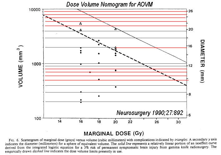
Stereotactic radiosurgery has been shown to treat successfully angiographically
demonstrated arteriovenous malformations of the brain. Angiographic obliteration has
represented cure and eliminated the risk of future hemorrhage. The role of radiosurgery in
the treatment of angiographically occult vascular malformations (AOVMs) has been
less well defined. In the initial 32 months of operation of the 201-source cobalt-60 gamma
knife at the University of Pittsburgh, 24 patients meeting strict criteria for
high-risk AOVMs were treated. Radiosurgery was used conservatively; each patient had
sustained two or more hemorrhages and had a magnetic resonance imaging-defined AOVM
located in a region of the brain where microsurgical removal was judged to pose an
excessive risk. Venous angiomas were excluded by performance of high-resolution
subtraction angiography in each patient. Fifteen malformations were in the medulla, pons,
and/or mesencephalon, and 5 were located in the thalamus or basal ganglia. Follow-up
ranged from 4 to 24 months. Nineteen patients either improved or remained clinically
stable and did not hemorrhage again during the follow-up interval. One patient
suffered another hemorrhage 7 months after radiosurgery. Five patients experienced
temporary worsening of pre-existing neurological deficits that suggested delayed
radiation injury. Magnetic resonance imaging demonstrated signal changes and edema
surrounding the radiosurgical target.
Dose-volume
guidelines for avoiding complications were constructed.
see dose nomogram) Our
initial experience indicates that stereotactic radiosurgery can be performed safely in
patients with small, well-circumscribed AOVMs located in deep, critical, or relatively
inaccessible cerebral locations.
Long-term results after stereotactic radiosurgery for patients with
cavernous malformations.
Hasegawa T, McInerney J, Kondziolka D, Lee JY, Flickinger JC, Lunsford LD.
Department of Neurological Surgery and the Center for Image-Guided Neurosurgery,
University of Pittsburgh, Pittsburgh, Pennsylvania 15213, USA.
Neurosurgery 2002 Jun;50(6):1190-7;
OBJECTIVE: Stereotactic radiosurgery has been used for patients with high-risk cavernous
malformations of the brain. We performed radiosurgery for patients with symptomatic,
imaging-confirmed hemorrhages for which resection was believed to be associated with high
risk. This study examines the long-term hemorrhage rate after radiosurgery. METHODS: We
reviewed data obtained before and after gamma knife radiosurgery on 82 patients
treated between 1987 and 2000. Most patients had multiple hemorrhages from brainstem or
diencephalic cavernous malformations. Follow-up data were examined to identify
hemorrhages, and an overall hemorrhage rate was calculated. RESULTS: Observation before
treatment averaged 4.33 years (range, 0.17-18 yr) for a total of 354 patient-years. During
this period, 202 hemorrhages were observed, for an annual hemorrhage rate of 33.9%,
excluding the first hemorrhage. Temporal clustering of hemorrhages was not significant.
After radiosurgery, patient follow-up averaged 5 years (range, 0.42-12.08 yr), for a total
of 401 patient-years. During this period, 19 hemorrhages were identified, 17 in the first
2 years posttreatment and 2 after 2 years. The annual hemorrhage rate was 12.3% per year
for the first 2 years after radiosurgery, followed by 0.76% per year from Years 2 to 12.
Eleven patients had new neurological symptoms without hemorrhage after radiosurgery
(13.4%). The symptoms were minor in six of these patients and temporary in five.
CONCLUSION: Radiosurgery confers a reduction in the risk of hemorrhage for high-risk
cavernous malformations. Risk reduction, although in evidence during initial
follow-up, is most pronounced after 2 years. Given the difficulty of identifying high-risk
patients, treatment after one major hemorrhage should be considered in selected younger
patients. Such a strategy warrants further investigation.
Treatment of symptomatic AOVMs with radiosurgery.
Kida Y, Kobayashi T, Tanaka T.
Department of Neurosurgery, Komaki City Hospital, Japan. Acta Neurochir Suppl (Wien)
1995;63:68-72
In spite of great success in the treatment cerebral AVMs with stereotactic radiosurgery,
the role of this treatment modality in angiographically occult vascular malformations
(AOVMs) is not recognized. Since the installation of the Gamma-knife, we have treated
20 cases of AOVMs by radiosurgery. There were 13 males and 7 females, the age ranged from
3 to 58 years with an average age of 34.0 years. Their clinical presentations at the onset
were haemorrhage in 11, convulsive seizure in 7 and progressive neurological deficits in
2. Two cases had multiple lesions. Among 20 symptomatic lesions, 14 were located
supratentorially, 4 in the brain stem and 2 in the cerebellar hemispheres. Following
localization with MRI and dose planning, the lesions were treated by radiosurgery and the doses
ranged from 15 to 20 Gy at the margins. Follow-up studies indicate a significant
control of rebleeding as well as of the convulsive seizure. Imaging studies demonstrated
the shrinkage of the lesion in 3 and reduced enhancement with Gadolinium-DTPA in some
others. Adverse effects, chiefly related to radiation-induced oedema, occurred in 5. But
they were generally mild and well controlled by medication. Thus the preliminary results
indicate a certain usefulness of radiosurgery in the treatment of symptomatic
AOVMs.
Radiosurgery of intracranial cavernous malformations.
Kim DG, Choe WJ, Paek SH, Chung HT, Kim IH, Han DH. Acta Neurochir (Wien)
2002 Sep;144(9):869-78
Department of Neurosurgery, Seoul National University College of Medicine, Clinical
Research Institute, Seoul National University Hospital, Seoul, Korea.
BACKGROUND: The efficacy of radiosurgery in cases of surgically high risk symptomatic
cavernous malformations (CMs) for reducing haemorrhagic risk and for seizure control has
not been clearly documented and the radiation-induced complications of radiosurgery remain
problematic. The authors present a retrospective clinical analysis of 22 cases of CMs
treated by radiosurgery. METHODS: Twenty-two patients with symptomatic CMs were treated by
linear accelerator (LINAC) radiosurgery or Gamma knife (GK) between 1995 and 1998. Medical
records including radiological investigations were carefully reviewed to the last
follow-up. The mean age of the patients was 34.1 years (12-56) and the male to female
ratio was 12:10. Twenty patients reported at least one episode of bleeding and four had
undergone microsurgery before radiosurgery. The remaining two patients presented with
seizure without evidence of recent haemorrhage. In 16 cases, the CMs were deep-seated, and
the others were located in the cerebral hemispheres; four were located at an eloquent
area. LINAC radiosurgery using computed tomography scan was performed in 11 cases until
May 1997, after which GK radiosurgery using magnetic resonance (MR) image was performed in
11 cases. The volume of the lesion ranged from 0.09 cc to 4.8 cc (mean 1.42 cc) and the
mean marginal dose was 16.1 Gy (8-24). The median follow-up period after radiosurgery was
38.3 months (21-67). The rate of haemorrhage, seizure, and neurological deterioration
following radiosurgery was analyzed, and the rate of haemorrhage was compared to that seen
in natural course reports. FINDINGS: There was one case of haemorrhage during the
follow-up period and the seizure was well controlled with anticonvulsants. In the group
with prior haemorrhage, the bleeding rate of cavernous malformation after radiosurgery
(1.55%/year) was lower than that of pre-radiosurgical period (35.5%/year, t=1.296,
P=0.04). Six patients showed neurological deterioration following radiosurgery, however,
the neurological deficits persisted in only two of the patients with LINAC. The
radiosurgical modality (LINAC vs. GK) showed a possible correlation to radiation induced
neurological deficits (P=0.06). On the MR images at the last follow-up, the lesion was
decreased in eleven patients, increased in one, and no change was found in 10 cases. The
T2 weighted MR images revealed a perilesional high signal change in nine patients. This
signal change was not statistically related to lesion size (P=0.236), location (P=0.658),
nor radiation dose (P=0.363), but was dependent on the treatment modality (P=0.02).
New-enhancing lesion and a new cyst were each found in one case, respectively, during the
follow-up. INTERPRETATION: Radiosurgery may be a good alternative option for
treatment of surgically high risk CMs. However, the optimal radiosurgical technique, dose
adjustment, and proper delineation of the mass are prerequisites. Radiosurgery induced
complications are still problematic and post-radiosurgery MR image changes need to be
further elucidated.
Radiosurgery for venous angiomas.
Lindquist C, Guo WY, Karlsson B, Steiner L. J Neurosurg 1993 Apr;78(4):531-6
Department of Neurosurgery, Karolinska Institute, Stockholm, Sweden.
Radiosurgical treatment with the gamma knife for venous angiomas was used as an
alternative to microsurgical removal in order to avoid abrupt cessation of venous
drainage, which may be shared by the venous angioma and important parts of the brain.
Thirteen cases of venous angioma were treated between 1977 and 1991. In two cases
cavernous angiomas were also present and in one case a distant arteriovenous malformation
(AVM) was also found. In two cases the angioma shared the venous drainage with an
adjoining AVM; this is the first description of such pathology. For venous angiomas
irradiation was prescribed to cover at least the convergence of the medullary veins. For
AVM's close to a venous angioma the treatment was exclusively prescribed to the AVM nidus.
After treatment, complete obliteration of the venous angioma was observed in one case,
partial obliteration was observed in three cases, and five venous angiomas were unaffected
by the treatment. Undue effects of radiation occurred in four cases: one focal edema and
three radionecroses. Extirpation of the radionecrotic tissue 6 months after radiosurgery
was necessary in one case. In the other three cases, the venous angioma was observed to be
completely or partially obliterated, or unaffected by the treatment (one case each). In
two cases of combined AVM and venous angioma, complete obliteration of the treatment AVM
nidus was obtained. It is concluded that radiosurgery for venous angioma, although
conceptually attractive, still does not fulfill the rigid criteria of minimal risk which
must be set for the treatment of a lesion with a benign natural history.
Gamma knife radiosurgery of the brain stem cavernomas.
Liscak R, Vladyka V, Simonova G, Vymazal J, Novotny J Jr. Minim
Invasive Neurosurg 2000 Dec;43(4):201-7
Department of Stereotactic and Radiation Neurosurgery, Hospital Na Homolce, Prague, Czech
Republic. Roman.Liscak@homolka.cz
Over 6 years (1992-1998) 26 patients with brain stem cavernomas were treated using the
Leksell gamma knife in Prague. 25 patients had a follow up of 6-66, median 24 months.
Annual risk of bleeding before radiosurgery was 4%. After gamma knife treatment sudden
impairment of neurodeficit reported as rebleeding was observed in 4 patients at 6-51
months, median 16.5 months, after radiosurgery. This represented a 6.8% risk of rebleeding
after radiosurgery, which is not significantly different from the risk before
radiosurgery. MRI or CT was performed in 24 patients 6-48, median 24, months after
radiosurgery. There were no signs of rebleeding in any of the patients, nor any increase
of the cavernoma. A decrease of cavernoma size was observed in 8 (33%) of patients.
Temporary collateral edema after radiosurgery was detected in 5 (21%) of patients 3-12,
median 11, months after radiosurgery. Neurodeficit was observed in 21 of 26 patients
before radiosurgery. Improvement of the neurodeficit was detected in 9 (43%) of them 6-36,
median 8, months after radiosurgery. Temporary morbidity caused by collateral edema or
rebleeding occurred in 7 patients (28%) and permanent morbidity remained in 2 patients
(8%). 2 patients died because of rebleeding 6 and 51 months after radiosurgery and the
third patient for unrelated reason. Radiosurgery of the brain stem cavernomas was
indicated when there was bleeding in the history or progressive neurodeficit and
microsurgery was considered too risky. Leksell gamma knife radiosurgery of cavernomas has
proved its low morbidity and zero mortality. In case of an insufficient effect of
radiosurgery, or if the protective effect from rebleeding comes too late, morbidity and
mortality can correspond to the natural course of the disease, as it was left without any
treatment.
Gamma knife radiosurgery for cavernous hemangiomas.
Zhang N, Pan L, Wang BJ, Wang EM, Dai JZ, Cai PW. J Neurosurg 2000
Dec;93 Suppl 3:74-7
Department of Neurosurgery, Shanghai Gamma Knife Hospital and Shanghai Huashan Hospital,
People's Republic of China. nanzhang@sina.com
OBJECT: The authors analyzed the outcome of 53 patients with cavernous hemangiomas who
underwent gamma knife radiosurgery (GKS) and evaluated the benefit of the treatment.
METHODS: From 1994 to 1995, 57 patients were treated with GKS for cavernous hemangiomas.
The mean margin dose to the lesions was 20.3 Gy (range 14.5-25.2 Gy) and the
prescription isodose was 50 to 80%. The mean follow-up period was 4.2 years. Four patients
were lost to follow up. In 18 of 28 patients whose chief complaint was seizures, there was
a decrease in seizure frequency. Five of 23 patients with hemorrhage suffered rebleeding 4
to 39 months after GKS. Seventeen patients in whom the hemangiomas were located at the
frontal or parietal lobe had neurological disability and in five this was severe. Two
patients underwent resection of their hemangioma after GKS. Three experienced visual
problems. Follow-up imaging demonstrated shrinkage of the lesion in 19 patients.
CONCLUSIONS: A higher margin dose (> 16 Gy) may be associated with a reduction in
the incidence of rebleeding after GKS. Higher dosage and severe brain edema after GKS
may decrease the frequency and intensity of seizures at least temporarily. Gamma knife
radiosurgery may play a role in protection against hemorrhage and in reduction of the rate
of seizure in selected cases with the appropriate dose. |

