 General Management
Surgery alone appears to be adequate
treatment for small, low-grade tumors confined to the
ethmoids in which negative surgical margins can be obtained.
An ethmoidomaxillary resection with or without orbital
sparing is usually necessary. This procedure is combined
with preoperative or postoperative irradiation. A complete
resection with preservation of vital structures is
achievable by using a craniofacial approach. The experience
from the University of Virginia, however, has yielded no
firm conclusions regarding whether craniofacial resection or
more conservative surgery could be performed in early-stage
disease.
Dias reported on 35 patients with
ENB treated with gross tumor resection through a transfacial
approach with postoperative RT in 11 patients, craniofacial
resection (CFR) and postoperative RT in seven, exclusive RT
in 14, CFR alone in one, and a combination of chemotherapy
and RT in two. Radiation therapy median dose was 48 Gy.
Analysis of survival showed that the Kadish classification
best predicted diseasefree survival. The presence of
regional and distant metastases adversely affected
prognosis. Craniofacial resection plus postoperative RT
provided a better 5-year disease free survival rate (86%)
compared with the other therapeutic options used. The 5-year
disease-specific survival rate was 64% and 43% for the low-
and high-grade tumors, respectively. Disease free survival
was 46% and 24% at 5 and 10 years, respectively. Overall
survival was 55% and 46% at 5 and 10 years of follow-up,
respectively. Aggressive multimodality therapeutic
strategies, particularly CFR and adjuvant RT, yielded the
best treatment outcome.
Early lesions involving the ethmoids with little or no bony destruction or nerve invasion can be treated adequately by high-energy (photon or electron) radiation therapy with good cosmetic and functional results. Those with more extensive local disease benefit from surgery and adjuvant irradiation, although some have spoken against combined surgery and radiation therapy because of complications. Patients with locally advanced disease or high-grade tumors should receive aggressive treatment with combined modalities, such as surgery, radiation therapy, and chemotherapy.
For advanced lesions, in which
disseminated disease is likely, chemotherapy may improve
tumor control and decrease the incidence of distant
metastases. A combination of thiotepa, cyclophosphamide,
doxorubicin, vincristine, nitrogen mustard, and actinomycin-D
has been used. Wieden reported complete tumor
regression and 2.7-year survival in a patient with extensive
olfactory esthesioneuroblastoma treated with a combination
of wide local excision, chemotherapy with cisplatin and
5-fluorouracil (5-FU), and irradiation (55.8 Gy). A
retrospective review of 10 patients with recurrent
esthesioneuroblastoma treated with chemotherapy at the Mayo
Clinic suggested that cisplatin-based chemotherapy is active
in advanced, high-grade tumors. Survival from initial
chemotherapy treatment was 44.5 months (range, 3 to 130
months) in patients with low-grade tumors and 26.5 months
(range, 2 to 67 months) in patients with high-grade tumors.
Treatment, which could be classified in 898 reported cases,
consisted of surgery alone in 24% (226 cases), radiation
therapy alone in 18.4% (165 cases), combined surgery and
radiation therapy in 43.2% (388 cases), chemotherapy in
13.2% (119 cases), and in 11 cases (1.2%) bone marrow
transplant. In the reported cases follow-up could be
evaluated in 477 cases, while in only 234 cases a 5-year
follow-up was done; on these 20.5% had surgery only, 11.1%
radiation therapy, and 68.4% combined surgery and radiation
therapy. The best survival rates were obtained by combined
therapy, 72.5% versus 62.5% with surgery alone and 53.8%
with radiation therapy
Elective Neck Treatment
Esthesioneuroblastoma has been shown to
metastasize to the neck and remote sites. Although the sites
of metastases are widely variable and often atypical, Olsen
reported cervical lymph nodes to be the most common site,
developing in 10/21 patients (48%) in their series. Beitler
found cervical lymph node metastases to be as common as
local recurrence. In a literature review of 110 patients by
Bailey , 24 patients (22%) had metastatic disease, with
cervical lymph nodes being the most common site. Davis
compiled a retrospective review of patients and found that
the cumulative cervical metastasis rate reached 27% (55/207
patients). In general, because of the low incidence of
cervical lymph node metastasis (≤10%) in early-stage
disease, elective irradiation of the neck or a dissection is
not indicated. However, in patients with Kadish stage C
disease, the cervical metastatic rate climbed to 44% (25/57
patients). As noted previously, Monroe ) observed cervical
node metastasis in 6/22 patients (27%), incidence similar to
that reported by other authors. In 11 patients they treated
with elective neck RT no recurrences were noted, in contrast
to 4/9 (44%) in patients not receiving elective neck RT.
Thus, with advanced-stage disease, cervical nodes should be
initially managed by irradiation, radical neck dissection,
or a combination of both
Radiation Therapy Techniques
A combination of photons and electrons
with anterior fields provides good coverage for limited
ethmoidal disease when the tumor is confined anteriorly.
Beam arrangement can be modified for disease extending into
the orbit or maxillary sinus. Obturator or bolus may be
needed postoperatively to compensate for tissue deficit.
When intracranial or posterior extension is present or tumor
has spread into the maxillary sinus, a pair of perpendicular
(anteroposterior and lateral) portals with wedges or two
lateral wedge fields in conjunction with an open anterior
photon field will give good coverage of the treatment volume
with the dose inhomogeneity around 10% to 20%. Incorporation
of a vertex field eliminates the high inhomogeneous dose
along the junction line of the conventional three-field
technique. Treatment techniques are similar to those
described for treatment of paranasal sinuses. The orbits can
be spared or treated as the degree of extension dictates.
Occasionally, an anterior electron beam field may be needed
to supplement low-dose areas. When the electron beam is used
over air cavities, some dosimetry problems result. Eye
blocks must be positioned precisely to avoid undesirable
side effects.
When combined therapy is used, preoperative doses of 45 Gy
and postoperative doses of 50 to 60 Gy are indicated,
depending on the status of the surgical margins. Doses of 65
to 70 Gy are delivered with irradiation alone in patients
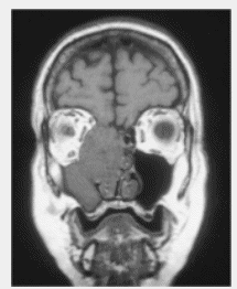
with inoperable tumors. Contrast-enhanced CT or MRI
scans before initiation of treatment are crucial to
demarcate extension of the tumor. Treatment planning with CT
for determination of tumor extension is extremely important.
Because of the proximity of esthesioneuroblastoma to the
optic nerves, optic chasm, and the brainstem, the precision
of treatment setup, target volume definition, and dose
homogeneity dictate tumor control and the sequelae of
treatment. Treatment techniques similar to those for
paranasal sinuses may create “hot spots” along the optic
tracks. High doses per fraction (exceeding 2 Gy) increase
the possibility of late sequelae such as blindness and bone
and brain necrosis.
|