In this
clinical study of IMRT aiming at
reducing dysphagia,
we have found statistically
significant, and potentially
clinically important,
dose–volume effect relationships
for dysphagia and aspiration,
which can serve as initial
dosimetric goals for IMRT. These
relationships support the
hypothesis that reducing the
doses to the swallowing
structures may reduce the
prevalence and severity of
dysphagia; however, they
do not yet prove this hypothesis
because they do not establish a
cause–effect association. In any
case, our findings motivate
efforts to further reduce these
doses, without compromising
target doses. The limiting
factor in this regard is the
percentage of the volume of each
of the swallowing structures
that is encompassed by the PTVs,
found in our study to correlate
highly with the mean doses to
the whole structure. The first
step in the efforts to improve
the sparing of the swallowing
(and other) structures in this
series has been made by daily
on-line imaging and correction
of setup deviations, which
facilitated reducing PTV margins
to 3 mm. Future efforts at our
institution include the
elimination of PTV margins and
the construction of IMRT plans
that cover the CTVs and their
known distribution of setup
uncertainties . Additional
potential strategies like proton
beam IMRT or structure and
target assessments and
adaptation during therapy should
be evaluated.
We have found significant
dose–volume effect relationships
regarding aspiration for the PCs
as a whole and also for each of
their parts: the superior,
middle, and inferior
constrictors.
These relationships were
statistically strongest for the
superior constrictor. The
importance of the superior PC
doses may be explained by the
details of the swallowing
mechanism. Elevation of the
larynx and pharynx during the
swallow is essential for airway
protection and bolus propulsion.
This elevation is facilitated by
the contraction of longitudinal
muscles (glossopharyngeus,
stylopharyngeus,
salpingopharyngeus, and
palatopharyngeus), which blend
with the circular fibers of the
superior constrictor As the
larynx and pharynx are pulled up
and forward by these muscles,
they are pulled away from the
lower posterior pharyngeal wall
and facilitate opening of the
upper esophageal sphincter at
the cricopharyngeus level. These
mechanisms of swallowing and
protection from aspiration, as
well as our VF-based results,
suggest that the benefits from
efforts to spare the swallowing
structures are likely to be
maximized if they include the
superior constrictors rather
than being confined to the
esophagus and its upper inlet.
Our findings that
patient-reported dysphagia was
also highly correlated with the
doses to the superior PC serve
as an independent validation of
the importance of sparing this
structure. In addition, a
recently presented study in
which brachytherapy was found to
reduce dysphagia, concluded that
the doses to the upper and
middle constrictors were the
most significant predictors of
patient-reported dysphagia.
We have also
found significant correlations
between the dose–volume
parameters in the GSL and
dysphagia. Several recently
presented studies examined
various dysphagia endpoints
after conventional radiotherapy
and
found significant correlations
with the doses to the
supraglottic or glottic larynx.
In general, these correlations
were similar to those reached by
our longitudinal study, in which
the endpoints were the
differences between the pre- and
the postradiation dysphagia
measures (rather than the
postradiation dysphagia alone).
In aggregate, these
studies affirm the potential
benefits in reducing the doses
to both glottic and supraglottic
larynx.
The
dose–volume effect relationships
for the swallowing structures
may depend on the intensity of
the chemo-RT regimen.
In the present study, no
strictures were observed in
patients receiving mean PC dose
<66 Gy. In comparison, we
have
previously found that after an
intensive gemcitabine-RT
regimen, the minimal dose
associated with strictures was
50 Gy. The differences
are likely related to the
severity of acute mucositis and
its consequential effect on
pharyngeal tissue.
Chemo-RT regimens that do not
differ markedly in the rate and
severity of the acute mucositis
seem to cause similar types and
rates of swallowing
abnormalities. We
therefore anticipate that the
dose–volume effect relationships
found in the present study,
which used a moderate-intensity
chemo-RT regimen, will be
reproduced after other commonly
used regimens of chemo-RT. The
site of the primary tumor also
affects dose–response
relationships, because different
primary tumor sites were found
to be associated with different
rates of both pre- and
posttherapy swallowing
abnormalities. The relative
homogeneity of the patient
population in our study, most of
whom had oropharyngeal cancer,
may have facilitated identifying
the dose–response relationships
for the swallowing structures.
The 3-months posttherapy
swallowing results reported
here, as well as the dose–volume
effect relationships, may change
over longer observation time.
Swallowing seems to reach a
steady state after approximately
12 months, as edema subsides and
long-term fibrosis develops
This issue will be addressed as
we continue to collect
swallowing endpoints at 12 and
24 months.
Swallowing-related laryngeal and
pharyngeal motion during
treatment may change dose
distributions in these
structures compared with those
observed in the simulation CT. A
detailed study of these effects
found that the incidence and
duration of swallowing during RT
is very low, averaging 0.45%
(range, 0–1.5%) of the total
irradiation time. Also, the mean
doses to the swallowing
structures were found in our
study to be highly correlated
with the percentages of the
structure volumes inside the
PTVs. In a previous study, we
found that these percentages did
not change significantly when
expansion of the swallowing
structures to produce planning
organ-at-risk volumes was made,
compared with the non-expanded
structures (5).
These data suggest that
expanding the swallowing
structures to obtain their
respective planning
organ-at-risk volumes would not
alter substantially the
planning, optimization, or
results of our study. This issue
deserves further investigation.
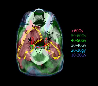
In conclusion, this study has demonstrated that IMRT aiming at sparing the swallowing structures is feasible. Significant relationships were found between dose–volume parameters for these structures and objective and subjective measures of swallowing dysfunction and dysphagia. These relationships can now serve to define optimization goals, and they motivate efforts to reduce these doses as much as possible. Longer follow-up is clearly necessary. Most importantly, care in the outlining of targets in the vicinity of these structures, avoiding target underdosing, and determining and reporting the locations of locoregional recurrences, are essential to ensure that the rates of local recurrences do not increase compared with the rates observed previously after IMRT.
|
|