Treatment planning
The PTV for all cases
included the prostate as defined by our prostate magnetic
resonance imaging (MRI) protocol, three-dimensionally
coregistered with prostate computed tomography (CT) imaging,
matching fiducial to fiducial, plus up to 2 cm of contiguous
seminal vesicle and a 2-mm volume expansion in all directions,
except posteriorly, where the prostate abutted the rectum. In
this region, the margin expansion was reduced to zero, justified
by CK system targeting accuracy and reports that prostate cancer
does not invade posteriorly in the midline beyond Denonvilliers'
fascia. Intermediate-risk patients had a 5-mm dorsolateral
prostate-to-PTV expansion to account for their increased risk
and potential distance of extracapsular extension near the
neurovascular bundle (NVB). Typically, the 2-mm margin expansion
used in patients with favorable prognosis split the NVB as
defined on T1-weighted gadolinium-enhanced MRI, whereas the 5-mm
expansion used for patients with intermediate prognosis fully
encompassed it This specific MRI sequence was selected to
provide prostate capsular definition, apical definition, and NVB
visualization while simultaneously creating a void around the
implanted gold fiducial markers that enables the most accurate
combination of prostate contouring and MRI-to-CT image
coregistration for treatment planning. Although T2-weighted MRI
gives detailed intraprostatic anatomic information, such as
dominant intraprostatic lesion location and transition zone vs.
peripheral zone delineation, the tendency of the gold fiducials
to disappear within T2 hypointense signal areas has precluded
making it a routine part of our CK SBRT treatment planning image
fusion procedure. The urethra was identified by insertion of a
Foley catheter, which also provided another reference structure
to use in MRI-to-CT image coregistration.
It is our hypothesis
that the CK may be used to deliver HDR-like dosimetry to the
prostate noninvasively. Supporting this hypothesis, our
dosimetry comparison between CK SBRT and simulated HDR showed
close similarities between them in coverage of the prostate PTV
by the prescribed radiation dose, as indicated by similar V100
characteristics. The CK SBRT also created a similar pattern of
dose escalation within the prostate peripheral zone compared
with HDR, although the absolute peripheral zone radiation dose
distribution was greater in the simulated HDR plans, reflecting
the physics inverse square law by which extreme radiation dosage
is created in immediate proximity to HDR source dwell positions.
Urethra sparing was
clearly more effectively accomplished by means of CK SBRT
in this study, with 29 of 30 comparisons favoring the CK SBRT
plans, typically by a dose difference on the order of 600 cGy.
In all except one case, attempts to match CK SBRT urethra dose
sparing with simulated HDR treatment plans resulted in deviation
in PTV V100 values for the HDR plans to less than the protocol
requirement of 95% PTV coverage. Thus, the CK SBRT plans
appeared to better maintain the protocol PTV V100 coverage
requirement while also respecting the urethra dose limit when
compared directly with case-matched, identically contoured,
simulated HDR brachytherapy treatment plans. To our knowledge,
this finding was not reported by other investigators and
therefore requires additional evaluation by other investigators
before definitive conclusions may be drawn.
A higher bladder Dmax
was obtained with simulated HDR plans, reflecting the
proximity of HDR point source dwell positions relative to the
bladder, whereas the higher bladder D10 level seen in the CK
SBRT plans likely reflects the effect of streaming CK radiation
beams through larger bladder volumes. The clinical significance
of these observed bladder dosimetry differences is unknown.
Nearly identical rectal
wall Dmax values were obtained with CK SBRT and HDR plans, with
a slightly lower median rectal mucosa Dmax value observed in CK
SBRT plans. With increasing distance from the point of
maximum rectal dose exposure (Dmax), progressively larger
differences in rectal wall and mucosa radiation dose sparing in
favor of CK SBRT were observed, indicating sharper dose falloff
beyond the rectal Dmax point with CK SBRT relative to HDR.
Because reported HDR rectal morbidity rates tend to be very low
it is unclear whether this more rapid rectal radiation dose
falloff with CK SBRT will bring an added clinical benefit,
although it suggests that the low rectal injury rates observed
with HDR should be equaled or even lower with CK SBRT. In this
context, it was reported that a greater incidence of Grade 2 or
higher rectal bleeding with HDR brachytherapy was obtained when
a larger volume of the rectum received low- to moderate-dose
radiation (10–50% of prescribed); this is the rectal exposure
range at which we observed the largest differential rectal
sparing with CK SBRT relative to HDR Again, it should be
emphasized that until our intermodality dosimetry findings are
evaluated by other investigators, any conclusions regarding the
relative rectal-sparing capability of CK SBRT vs. HDR
brachytherapy are preliminary.
It should be noted
that both CK and HDR radiation dose sculpting platforms are
extremely user programmable, which complicates the direct
comparison of these radiation delivery modalities. It is
possible that some untested combination of HDR brachytherapy
catheter configuration and source dwell position instructions
would create a more favorable HDR brachytherapy result, although
our relative CK SBRT vs. HDR dosimetry observation trends seemed
consistent across the range of prostate volumes and catheter
configurations analyzed in our study. Likewise, it also is
possible that more effective CK SBRT treatment planning and
radiation beam collimator selection could create a more
favorable CK SBRT dosimetry result. Despite these caveats, the
present study clearly shows that treatment plans that closely
approximate those used in HDR brachytherapy for patients with
prostate cancer may be constructed and delivered using the CK
system.
Further intraprostatic
CK SBRT dose escalation
Our early
observation of dosimetry trends that favored HDR for the
volume of PTV exceeding the prescription dose by 25% or more
and CK SBRT for urethra sparing prompted us to run second CK
SBRT plan iterations in the first 5 patients. For this
exercise, the CK planning computer was instructed to match
the urethra Dmax of the corresponding HDR plan while
relaxing the CK PTV Dmax limitation and maintaining all
other dose limitations to see whether this approach would
allow CK SBRT plans to more closely resemble HDR V125 and
V150 values. This is exactly what we found; second-iteration
CK SBRT V125 and V150 measurements much more closely
approached the median simulated HDR V125 and V150 values,
more effectively matching HDR PTV dose escalation volumes.
Bladder Dmax and all rectal dosimetry parameters changed
very little in the second-iteration CK SBRT plans compared
with the de novo SBRT plans, although there was a more
significant and variable increase in the bladder D10
dosimetry statistic, indicating that more stringent
attention to bladder sparing may be required if extreme
intraprostatic dose escalation is attempted with CK.
In summary, our
second-iteration CK SBRT plans show that intraprostatic dose
escalation approaching parity with HDR appears possible,
although with attendant partial or complete loss of the
superior urethra sparing observed in the initial CK SBRT
plans. Because urethral strictures were reported with HDR
brachytherapy a reasonable strategy might be to escalate
intraprostatic dose as much as possible with CK SBRT while
still respecting the urethra dose limits of the CK protocol.
This is the approach we continue to use in our ongoing
Virtual HDRsm CyberKnife
clinical trial, which shows increasing PTV V125 and V150
trend lines over sequential patients.
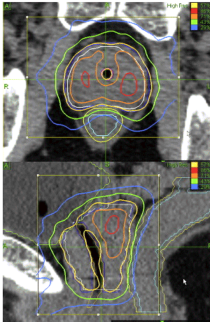
Although our CT
and MRI planning sequences are not specifically designed to
delineate the peripheral zone, inspection of the 125%
(orange) and 150% (red) isodose lines shown in
compared with the pathologic illustration suggests a
significant degree of coincidence between CK dose escalation
zones and the anatomic peripheral zone of the prostate. A
more direct approach would be to perform specific peripheral
zone and dominant intraprostatic lesion dosimetry
comparisons between the modalities from T2-weighted MRI
imaging sequences should such images become available for
inspection in future patients.
Radiobiologic relevance
of intraprostatic dose escalation
Whether the
radiation dose delivery platform is CK SBRT or HDR,
the prescription
dose of 38 Gy in four fractions is the dose calculated to
deliver a biologically lethal blow to the cancer; therefore,
it is unclear to what extent further dose escalation beyond
this level within the prostate may be necessary or
beneficial. However, the exact α/β ratio of prostate
cancer upon which hypofractionation schedules are calculated
remains uncertain, as discussed in the recent report of
Williams. If we use the HDR literature to justify CK
treatment, an argument may be made that CK practitioners are
well advised to mimic HDR intraprostatic dose distribution
as closely as possible to maximize the possibility of
reproducing the favorable HDR clinical result.
Intraprostatic dose escalation beyond the prescribed dose
level, as naturally occurs with any form of brachytherapy,
provides backup cancer-cell–killing power in the event that
cancer-cell populations with higher α/β ratios exist within
heterogeneous populations of prostate cancers and patients
with prostate cancer. It also should be noted that Lotan
obtained the most reliable tumor ablation in prostate
cancers in a nude mouse model with a dose of 45 Gy in three
fractions, a more aggressive dosing schedule than described
in our study or reported in the HDR literature, again making
a case for intraprostatic dose escalation.
Treatment delivery and
dosimetry accuracy
Even infinite
computerized dose-sculpting capability is clinically
meaningless unless the targeting accuracy of the delivery
system is sufficient to ensure precise delivery of the
treatment plan. Unlike typical image-guided radiotherapy
systems, which detect and correct the target volume position
only once at the beginning of each treatment, the CK robotic
delivery system uses a unique stereoscopic X-ray–based
tracking system that updates and corrects robotic linear
accelerator position with regard to both translational and
rotational target volume movements up to 100 or more times
per treatment, resulting in submillimeter targeting accuracy
for brain and spine applications
Because the
fiducial-based CK tracking procedure for prostate treatment
is comparable, the delivery accuracy for prostate cancer
theoretically should be identical to that described for
brain and spine applications, but with the important caveat
that prostate motion is potentially more complex to
accurately track and correct for than relatively more fixed
brain and spinal targets. Movement caused by bowel
peristalsis and bladder filling can cause a rapidly
shifting, rotating, and even deforming prostate target
volume Until the quantitative effects of these added
prostate motion and potential deformation complexities are
understood in greater detail, this remains a valid point of
criticism for investigators who discuss CK system targeting
accuracy in the treatment of prostate cancer.
HDR brachytherapy
accuracy is also subject to potential error and distortion
because of such factors as variable and potentially
significant HDR source transit dose contribution that is
neglected by HDR planning computers, prostate volume
fluctuation caused by needle trauma, and longitudinal HDR
catheter translocation during the course of the patient's
hospitalization, with the latter likely representing the
largest source of potential HDR brachytherapy targeting
error.
Regarding the
resemblance of the computer-generated treatment plan to the
actual delivered treatment, both CK and HDR brachytherapy
have potential dosimetry deviations. Because the sources of
dosimetry errors and targeting inaccuracy are different
between these modalities, their relative magnitudes are
speculative, and as such, it is unknown which modality most
consistently delivers its treatment plan more accurately.
Clinical discussion
Our Virtual HDRsm
CyberKnife clinical series is small, with maximum follow-up
limited to 12 months; however, some preliminary clinical
observations may be reported. Our
continuously
decreasing median 4-month post-CK PSA value of 0.95 ng/ml
suggests a similar response slope to that described by the
Stanford group, who reported a median 18-month post-CK PSA
value of 0.22 ng/ml. On a larger scale of comparison, our
observed median 4-month post-CK PSA decrease of 86% appears
comparable to the short-term PSA response magnitude reported
with other radiation-based approaches, including
standard external beam radiotherapy and
103Pd seed brachytherapy,
with much longer term follow-up required to assess
durability of the response. Early post-HDR brachytherapy PSA
responses, similar to the present data, have not been
reported to our knowledge. As our series matures, relative
PSA-based disease-free survival rates will be compared.
Acute toxicity of
Virtual HDRsm CK
monotherapy was self-limited and manageable, primarily
consisting of several months of α-blocker–dependent
irritative/obstructive uropathy, as well as fatigue, and a
less than 100% incidence of typically Grade I proctalgia/rectal
urgency that usually resolved by 3–4 weeks after CK
treatment. Although our early post-CK toxicity data
appear very encouraging, definitive toxicity assessment
requires significantly longer follow-up because serious
radiation-related complications may not manifest until 1 to
2 years posttreatment. Our series is too small and follow-up
is too short to make a meaningful statement about the
incidence of post-CK erectile dysfunction, although this
domain will be assessed in detail as the fully accrued study
matures. For long-term toxicity evaluation, the Virtual HDRsm
CK monotherapy protocol includes the long-form Expanded
Prostate Cancer Index Composite assessment, which measures
urinary, gastrointestinal, sexual, and hormonal-mediated
sequelae of therapy
Results
Planning
target volume coverage by the prescription dose was similar for
CK SBRT and HDR plans, whereas percent of volume of interest
receiving 125% of prescribed radiation dose (V125) and V150
values were higher for HDR, reflecting higher doses near HDR
source dwell positions. Urethra dose comparisons were lower for
CK SBRT in 9 of 10 cases, suggesting that CK SBRT may more
effectively limit urethra dose. Bladder maximum point doses were
higher with HDR, but bladder dose falloff beyond the maximum
dose region was more rapid with HDR. Maximum rectal wall doses
were similar, but CK SBRT created sharper rectal dose falloff
beyond the maximum dose region. Second CK SBRT plans,
constructed by equating urethra radiation dose received by point
of maximum exposure of volume of interest to the HDR plan,
significantly increased V125 and V150. Clinically, 4-month
post–CK SBRT median prostate-specific antigen levels decreased
86% from baseline. Acute toxicity was primarily urologic and
returned to baseline by 2 months. Acute rectal morbidity was
minimal and transient.