Stereotactic Radiosurgery Center, S. Bortolo
City Hospital, Vicenza, Italy.
federico.colombo@ulssvicenza.it
To
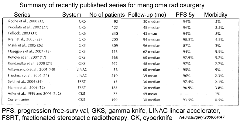
present initial, short-term results obtained
with an image-guided radiosurgery apparatus
(CyberKnife; Accuray, Inc., Sunnyvale, CA)
in a series of
199
benign intracranial meningiomas.
Selection criteria included lesions
unsuitable for surgery and/or remnants after
partial surgical removal. All patients were
either symptomatic and/or harboring growing
tumors. Ninety-nine tumors involved the
cavernous sinus; 28 were in the posterior
fossa, petrous bone, or clivus; and 29 were
in contact with anterior optic pathways.
Twenty-two tumors involved the convexity,
and 21 involved the falx or tentorium. One
hundred fourteen patients had undergone some
kind of surgical removal before
radiosurgery.
Tumor volumes varied from 0.1 to 64 mL
(mean, 7.5 mL) and radiation
doses ranged from 12 to 25 Gy (mean, 18.5 Gy).
Treatment isodoses varied from 70 to 90%. In
150 patients with
lesions larger than 8 mL and/or with tumors
situated close to critical structures, the
dose was delivered in 2 to 5 daily fractions.
Single session was used for small tumors at
least 3mm from the brainstem or optic
pathways, large tumors or closer were given
in 2-5 fractions with doses equivalent to
11-12Gy in a single session assuming an
alpha/beta of 3 for meningioma, tried to
reduce the dose absorbed by any portion of
the anterior optic pathways to less than 7Gy
per session. Also for large lesions (larges
than 3cm or 13.5cc volume) used
fractionation
| No of
fractions |
No of
Patients |
Total Dose
(Gy) |
BED (a/b/ 2) |
BED (a/b 3) |
| 1 |
49 |
11-13 |
71.5 - 97.5 |
51.3-69.3 |
| 2 |
32 |
14-17 |
63-89.2 |
46.6-65.1 |
| 3 |
76 |
16-20 |
58.4-86.6 |
44.2-64 |
| 4 |
18 |
18-23 |
58.8-89.1 |
45-67.8 |
| 5 |
24 |
19-25 |
64.1-87.5 |
49-66.7 |
RESULTS: The follow-up periods ranged from 1
to 59 months (mean, 30 months; median, 30
months). The tumor volume decreased in 36
patients, was unchanged in 148 patients, and
increased in 7 patients. Three patients
underwent repeated radiosurgery, and 4
underwent operations. One hundred fifty-four
patients were clinically stable. In 30
patients, a significant improvement of
clinical symptoms was obtained. In 7
patients, neurological deterioration was
observed (new cranial deficits in 2,
worsened diplopia in 2, visual field
reduction in 2, and worsened headache in 2).
CONCLUSION: The introduction of the
CyberKnife extended the indication to 63
patients (>30%) who could not have been
treated by single-session radiosurgical
techniques. The procedure proved to be safe.
Clinical improvement seems to be more
frequently observed with the CyberKnife than
in our previous linear accelerator
experience.
