|
|
|
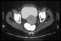
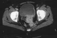
| Stage Ib carcinoma confined to the cervix. CT image shows a mass with slightly
heterogeneous area of attenuation expanding the cervix and surrounded by a thin rim of
relatively preserved stroma. The cervical margins are smooth, well defined, and intact.
Parametrial soft-tissue stranding or masses are lacking, and the periureteral fat planes
are preserved. |
|
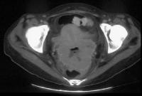
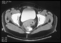
Cervix, cancer. CT image depicts a large lobulated mass replacing the cervix and showing non-uniform
hypoattenuation. The air and fluid in the center of the mass are consistent with tumor
necrosis and complicating infection (the patient had purulent discharge). The central
hypoattenuation in the uterine corpus is suggestive of minimal fluid in the cavity. |
|
|
|
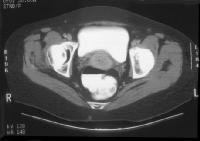
| Cervix, cancer. Clinical stage IIb
cervical carcinoma . The parametrial invasion is depicted
with CT as loss of definition of the cervical contours accompanied with increased
attenuation and prominent soft-tissue stranding in the parametrial fat. Parametritis can
result in similar findings. The cervix shows ill-defined hypoattenuation, but the tumor is
not clearly delineated. |
|
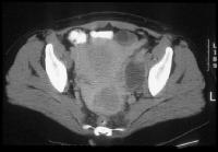
Cervix, cancer. CT demonstrates a
markedly enlarged lymph node at the left pelvic sidewall. This finding is consistent with
pelvic lymph node metastasis, which is indicative of stage
IIIb disease. The cystic consistency is not unusual for metastatic cervical carcinoma. The
primary tumor is well depicted as a hypoattenuating, circumscribed mass. A cyst is present
in the anteriorly located left ovary. |
|
|
|
| Cervix, cancer. CT demonstrates a
cervical tumor directly extending into the posterior wall of the bladder
and into the left pelvic sidewall. Extension into the pelvic sidewall is a feature of
stage IIIb disease whereas involvement of the bladder wall is a feature of stage IVa
disease. |
|
Cervix, cancer. Parametrial and rectal invasion by cervical carcinoma are depicted with CT as loss
of definition of the cervical contours accompanied with masslike soft tissue that replaces
the parametrial fat on the right and that extends into the anterior and right-sided rectal
walls. |
|
|
|
|
|
|