Radiation
Dose–Volume Effects in the Brain
IJROBP
We have reviewed the published data regarding
radiotherapy (RT)-induced brain injury.
Radiation necrosis
appears a median of 1–2 years after RT; however,
cognitive decline develops over many years. The incidence
and severity is dose and volume dependent and can also be
increased by chemotherapy, age, diabetes, and spatial
factors. For
fractionated RT with a fraction size of <2.5 Gy, an
incidence of radiation necrosis of 5% and 10% is predicted
to occur at a biologically effective dose of 120 Gy
(range, 100–140) and
150 Gy (range, 140–170), respectively.
For twice-daily fractionation, a steep increase in toxicity
appears to occur when the biologically effective dose is >80
Gy. For large fraction sizes (≥2.5 Gy), the incidence and
severity of toxicity is unpredictable.
For single fraction
radiosurgery, a clear correlation has been demonstrated
between the target size and the risk of adverse events.
Substantial variation among different centers'
reported outcomes have prevented us from making
toxicity–risk predictions. Cognitive dysfunction in children
is largely seen for whole brain doses of ≥18 Gy. No
substantial evidence has shown that RT induces irreversible
cognitive decline in adults within 4 years of RT.
|
| For radiosurgery, the incidence of necrosis
depends on the dose, volume, and region irradiated. The Radiation
Therapy Oncology Group conducted a dose-escalation study that sought
to define the maximal dose for targets of different sizes; all
subjects had previously undergone whole brain irradiation. The
maximal tolerated dose for targets 31–40 mm in diameter was 15 Gy,
and for targets 21–30 mm in diameter, it was 18 Gy. For targets <20
mm, the maximal tolerated dose was >24 Gy . The volume of brain
receiving ≥12 Gy has been shown to correlate with both the incidence
of radiation necrosis and asymptomatic radiologic changes. The large
variation in absolute complication rates among studies is difficult
to comprehend, but it might relate to differences in the definitions
of the volume and toxicity, the avoidance of critical structures,
and the type and length of clinical follow-up. |
|
 |
|
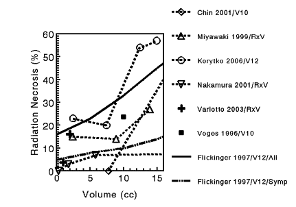
Relationship between volume
receiving high-dose irradiation and incidence of radiation necrosis
in single-fraction stereotactic radiosurgery. Studies differed in
their completeness of follow-up, definition of volume, and
definition of radiation necrosis. Graph based on data presented.
Volume plotted as a point, representing mid-point of volume range. V10
= volume receiving 10 Gy; V12 =
volume receiving 12 Gy; RxV = treatment volume. Flickinger data is
shown for patients with either radiologic or symptomatic evidence of
necrosis (marked as "All"), or only those with symptomatic necrosis
(Symp). The other authors' data refers to symptomatic necrosis.
|
| For fractionated RT, the relationship between
the radiation dose and radiation necrosis for partial brain
irradiation is presented segregated by the fractionation scheme.
Different fractionation schemes were compared using the biologically
effective dose (BED) with an α/β ratio of 3.
For standard fractionation,
a dose–response relationship exists, such that an incidence of side
effects of 5% and 10% occur at a BED of 120 Gy (range, 100–140) and
150 Gy (range, 140–170), respectively (corresponding to 72 Gy
[range, 60–84] and 90 Gy [range, 84–102] in 2-Gy fractions).
For twice-daily fractionation, a steep increase in toxicity appears
to occur when the BED is >80 Gy. For daily large fraction sizes
(>2.5 Gy), the incidence and severity of toxicity is unpredictable.
The reader is cautioned against overinterpreting the data presented,
which was created from a heterogeneous data pool (i.e., different
target volumes, endpoints, sample sizes, and brain regions). No
evidence has shown that children are especially at risk of radiation
necrosis |
|
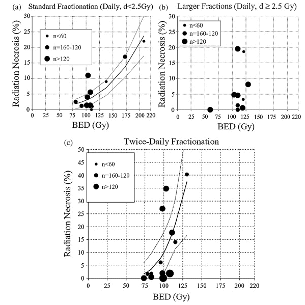 |
| Relationship between biologically effective
dose (BED) and radiation necrosis after fractionated radiotherapy.
Fit was done using nonlinear least-squares algorithm using Matlab
software (The MathWorks, Natick, MA). Nonlinear function chosen was
probit model (similar functional form to Lyman model). Dotted lines
represent 95% confidence levels; each dot represents data from
specific study, n = patient numbers as shown. (a) Fraction size <2.5
Gy; (b) fraction size ≥2.5 Gy (data too scattered to allow plotting
of “best-fit” line); and (c) twice-daily radiotherapy. |