| In patients who have brain
metastases that impinge on eloquent areas, or are too large,
numerous, or disseminated for surgery or RS,
Whole-Brain Radiotherapy (WBRT) alone remains the treatment of
choice and provides effective symptom relief in the
majority of cases. Although response rates after WBRT vary,
complete or partial responses
have been documented in more than 60% of patients in
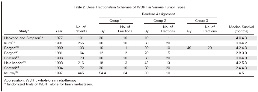
randomized controlled studies conducted by the RTOG. Table
summarizes results of different dose and fractionation
schedules from eight randomized studies in patients with brain
metastases who received WBRT alone, with
median survival ranges from 2.4 to
4.8 months. The consensus from these studies of
fractionation schedules is that differences in dose, timing,
and fractionation have not significantly altered the median
survival time for WBRT treatment of brain metastases.
Currently, the most common WBRT
regimen uses 30 Gy in ten 3 Gy fractions. Though this dose is
clearly inadequate for long-term tumor control, except in the
most radiosensitive histologies such as germinoma and lymphoma,
this lower dose has traditionally been used to minimize the
toxicity of WBRT. An analysis of RTOG trials shows that this
results in control of
disease in approximately 50% of patients at 6 months.
A
second major use of WBRT is as an adjuvant after surgery or
radiosurgery. MRI reveals that approximately 80% of patients
have more than one metastasis, and approximately 50% have three
or more metastases. Moreover, 70% of patients with brain metastases
experience relapse after resection, if WBRT is omitted.
CONTROVERSY SURROUNDING THE USE OF WBRT WITH
RADIOSURGERY
Radiosurgery after WBRT has been validated
with level 1 evidence as a standard of care option in the
management of patients with single brain metastases.
As radiosurgery
has increased in popularity, a new trend has been emerging in
the management of patients with brain metastases; in this
approach, patients with a limited number of brain metastatic
lesions (the exact definition of limited is based on
institutional preference and varies from three to ten or more)
are treated with radiosurgery without WBRT, and are then
closely monitored, which involves monthly or every other month
MRIs. Repeat radiosurgery, is performed for new intracranial
metastases, with the intent being avoidance or delay of WBRT in
as many patients, and for as long as possible. The putative
rationale for this is the avoidance of significant
neurotoxicity from WBRT; unfortunately, this approach has not
been validated in controlled clinical trials, and its application
dramatically increases the overall cost of managing these patients,
with multiple and expensive imaging studies (total charges of
easily $2,000 per study or more) and repeat radiosurgical procedures
(often $20,000 to $40,000 total charge per procedure).
In this section, we will discuss the
elements that drive this debate. We summarize the arguments as
follows: (1) most patients have oligometastatic disease; (2)
survival is unaltered whether upfront WBRT is used or not; (3)
radiosurgery is good enough for local control; and (4) the
neurologic status and quality of life of patients in whom WBRT
is withheld is superior.
A proposition in support of withholding
WBRT is that most patients have oligometastatic disease. (The
term 'oligometastasis' was initially used to describe a restricted
locoregional tumour load, but the term has now become synonymous with
isolated distant metastases.) Older
autopsy and computed tomography (CT) imaging studies suggest
that the rate of multiple brain metastases ranges from 58% to
86%, with a mean of 66%, but these studies have been criticized
on several grounds. Few prospective brain metastases trials
have mandated routine MRIs, and therefore these data are
sparse. In one MRI-based study, only 19% of the 336 patients
had a single lesion; the percentage of patients with two,
three, four, and five or more lesions was 16%, 13%, 10% and
40%, respectively. In most trials
of radiosurgery, three is considered the upper limit in terms
of the definition of oligometastases, and in this trial,
50% of patients had more than three lesions on their MRIs.
Therefore, most
reports would suggest that only approximately 20% of patients
have one brain metastasis, and this is especially important, as
evidence-based data suggest no survival benefit from aggressive
local treatments, such as surgery or radiosurgery in patients
with more than one metastatic lesion.
A second contention is that survival is
unaltered whether upfront WBRT is used or not. In a disease
process where the occurrence of brain metastases represents
only one component of systemic spread, it is unlikely that
local control of disease in one compartment will alter overall
survival, and decisions regarding local disease control are not
driven by the impact on survival, but rather on the value of
local control.
Not surprisingly, trials (that were not designed to
answer an overall survival question) of local therapies that
have excluded WBRT show no difference in overall survival.
However, a cautionary observation is provided
by a retrospective analysis from Germany; Pirzkall et al
reported that for brain metastases
patients without extracranial disease, (ie, patients with a
much lower likelihood of dying from systemic metastases) the
median survival after radiosurgery alone with WBRT used for
salvage was 8.3 months compared with 15.4 months for similar
patients treated up front with radiosurgery plus WBRT.
Similar results were seen in a retrospective study
from the Mayo Clinic with a survival benefit for adjuvant WBRT
limited to patients without systemic disease—5-year
survival rates of 21% for those who received adjuvant WBRT
compared to 4% for those patients who did not.
These observations are crucial, implying that
for those patients where prolonged survival is likely, failure
to control the intracranial disease by omitting or delaying
WBRT could potentially result in a negative survival impact.
It has been proposed, primarily based on
retrospective institutional chart reviews, that radiosurgery is
good enough for local control. In the prospective randomized
Japanese trial, JROSG 99-1, patients were randomly assigned to
radiosurgery alone, versus WBRT and radiosurgery. The
actuarial 6 month freedom from new
brain metastases was 48% in the radiosurgery alone arm, and 82%
in the radiosurgery and WBRT arm (logrank, P = .003).
Actuarial 1 year brain tumor control rate for the lesions
treated with radiosurgery was 70% in the radiosurgery alone arm
and 86% in the radiosurgery and WBRT arm (log-rank, P = .019).
Another randomized trial compared radiosurgery alone, WBRT
alone, or WBRT and radiosurgery. The local brain control rate
was highest in the radiosurgery plus WBRT arm. A prospective
single arm, multi-institutional ECOG phase II study of
radiosurgery alone for radioresistant histologies (melanoma,
sarcoma, renal cell carcinoma) in patients with one to three
brain metastases has recently been reported.
At 6 months, 39.2% failed within the
radiosurgery volume and 39.4% failed outside the radiosurgery
volume, thereby defining the limited benefit from radiosurgery
alone. Clinical trial-based assessments therefore suggest high
rates of intracranial failures and reduced local control rates
when WBRT is omitted or delayed.
It has been speculated that the neurologic
status and quality of life of patients in whom WBRT is withheld
is superior. The only randomized data available in this context
are from a recent Japanese trial, not yet fully published. In
that study, patients were randomly assigned to radiosurgery
alone or with WBRT; detailed neurocognitive assessments were
not performed, and the primary assessment was by an evaluation
of performance score and neurologic functional status using
RTOG criteria. There were no
differences in these end points between the two study arms,
belying the claims of worse neurologic outcomes in the WBRT arm.
In fact, many have argued that the converse
might be true, (ie, withholding WBRT increases intracranial
failure and neurologic deterioration is more directly related
to disease progression in the brain.) In a recent phase III
trial of WBRT with or without the radiosensitizer, MGd, the
most significant predictor for neurologic and neurocognitive
decline, as well as deterioration in quality of life was disease
progression in the brain.
Therefore, the switch to omitting WBRT has
largely been made in the absence of prospective randomized data
and needs to be considered carefully. In large measure, it is
fair to say that this switch has been made because physicians
have observed some patients experiencing neurocognitive and
neurologic decline (the causes for which could in fact be
multifactorial).
NEUROCOGNITIVE AND NEUROLOGIC DECLINE
Neurocognitive Function: Baseline, Post-WBRT, and Predictive Variables.
Patients with brain metastasis may suffer a certain degree of
neurocognitive function (NCF) impairment from multiple factors
including the tumor, WBRT, neurosurgical procedures, chemotherapy
and other neurotoxic therapies (including anticonvulsants and
steroids), or from paraneoplastic effects induced by the malignancy.
Furthermore, radiotherapy variables (eg, total
dose, volume of brain irradiated, fraction size) and the
interaction with other treatment (eg, concurrent chemotherapy)
or patient variables (ie, age, diabetes mellitus) all influence
the incidence of radiation-induced injury to the brain and may
account for the differences in reported incidences of cognitive
deficits.
For example,
two studies have not found
significant cognitive decline if whole brain fraction size was
less than 3 Gy.
In addition, in a phase III clinical
trial that compared WBRT (30 Gy in 10 fraction) versus WBRT
plus MGd in patients with brain metastasis, analysis of NCF
data demonstrated that 90.5% of
patients had significant impairment (>1.5 SDs from the age-adjusted
population normalized score) in one or more neurocognitive domain
at the time of diagnosis of brain metastasis, with 42% of the
patients having impairment in at least four out of the eight
tests. This result is consistent with previous reports
where baseline neurocognitive and neuropsychological test scores
were impaired in patients with small-cell lung cancer (even
in the absence of overt brain metastases) before receiving WBRT.
On reviewing NCF data from patients with
brain metastases who received WBRT alone, we found that at 3
months, the mean NCF test scores appear to be unchanged or
slightly reduced. However, 3 months later, gradual recovery is
observed . To determine if this recovery is due to patient
selection effect, we compared groups of patients who were alive
at 4 months after treatment with patients who were alive at 15
months after treatment. We found that 4-month survivors had a
sharp drop in their mean NCF scores in the first few months,
whereas 15-month survivors had stable NCF with gradual
improvement over time. Although both groups of patients had
reduction in mean tumor volume from WBRT in the first 4 months,
greater reduction was observed in the long-term survivors,
which may explain their relatively stable NCF (J. Li,
manuscript in preparation). These
findings are consistent with a previous report that NCF decline
correlates with tumor growth. In addition, these results
suggest that the initial changes in patients' NCF test scores,
as well as radiologic evidence of tumor regression, may be
predictive of overall survival. Although it is possible that
the improvement in NCF scores in long-term survivors may also
result from increased familiarity with retesting, the battery
of NCF tests utilized in this study was designed to have
minimal effect from repetitive testing.
A more definitive predictor of survival in
patients with brain metastasis is baseline NCF. Univariate
analysis has shown that all three domains of NCF testing
(memory, fine motor, executive function) at baseline are
predictive of survival, whereas multivariate analysis showed
that only memory score was an independent predictor.
Similar results were obtained in patients with
recurrent brain tumors, where memory test score was found to be
highly related to survival by multivariate analysis.
Interestingly in this patient population,
cognitive deterioration occurred almost 6 weeks before
radiographic failure, highlighting the sensitivity and survival
predictive value of NCF changes.
Our preliminary analysis of patients with brain
metastasis treated with WBRT has also shown that certain
functions may deteriorate earlier in the course of the disease
than others. If confirmed, these results may help to further
streamline NCF tests that are more sensitive in predicting
subsequent QOL changes and survival.
Studies involving NCF deterioration should
be interpreted cautiously. NCF decline in the literature is
often defined statistically and there is little consensus as to
the actual clinical relevance of a statistical definition. Use
of different definitions may artificially change the sequence
of events, suggesting a further need for a statistical model
that allows use of early test scores to predict later events
without requiring such predefined statistical windows. In
addition, conventionally used measures such as the Folstein
mini-mental examination (MMSE) are rather crude and it is
crucial to develop sensitive and practical NCF testing to
characterize these changes. In particular, the sensitivity of
MMSE has been shown to be problematic in detecting subtle
neurocognitive dysfunction
in patients with brain metastasis where
clinically apparent WBRT-induced
dementia is rare (1.9 to 5.1%).
Recent evidence indicates that a battery of validated,
language-specific, and population-normalized NCF tests evaluating
memory, fine motor coordination, and executive functions confers
more accurate and comprehensive measurement of NCF changes in
patients with brain metastasis.
Correlation Between NCF and Quality of Life
Although neurocognitive function is believed to have a major
impact on the quality of life (QOL) in patients with many medical,
mental, or psychiatric diseases
this relationship has not been adequately
evaluated in patients with brain metastases. An early study in
patients with primary brain tumors did show that NCF impairment
caused a decline in functional independence more often than
physical disability. This suggests that any treatment that
would reduce the severity of or prolong the time to NCF
deterioration could lead to increased functional status in
these patients.
The recent development of brief, comprehensive,
and validated measurements for NCF and cancer-specific QOL has
made it possible to explore the nature of the relationship between
NCF and functional performance in anticancer clinical trials.
In patients with brain metastasis who
received WBRT alone,
our analysis has shown that at baseline each
individual NCF test score is correlated with functional
measures such as FACT-Br (Functional Assessment of Cancer
Therapy–Brain, brain-specific QOL), Barthel Index, or KPS. The
strength of these correlations ranges from weak to moderate,
but all are statistically significant. Interestingly, the
correlations became stronger 4 months after treatment, and the
highest correlation coefficient is observed between memory and
Barthel Index. The correlations remain strong at 6 months
although statistical significance diminishes due to patient
loss (J. Li, manuscript in preparation). These results clearly
demonstrate that there is an association between NCF and QOL in
patients with brain metastasis. In addition, these results
suggest that therapy that preserves patients' NCF, especially
memory, may have a positive impact on patients' quality of
life.
The sensitivity of the Barthel index and
FACT-Br for detecting decline in functional status in patients
with brain metastasis or primary brain tumors has been
questioned. In fact, it appears from the literature, that gross
decline in QOL, especially in terms of severe impairment in
activities of daily living may indeed be a late event. Even
with the relative insensitivity of current QOL tools, in
patients with brain metastasis receiving WBRT, we consistently
detected a correlation between NCF and QOL, suggesting that
more sensitive QOL testing could help potentially delineate
changes in patient's functional status at an earlier point in
time.
Mechanism of Neurocognitive
Dysfunction
The response to radiotherapy has been classically divided into
three categories based on the timing of onset of symptoms: acute,
subacute, and late.
Acute effects
occur during the first few weeks of treatment and are often
characterized by drowsiness, headache, nausea, vomiting, and
worsening focal deficits. Often, cerebral edema is the cause of
these symptoms and corticosteroids may improve these symptoms.
Subacute encephalopathy
(early delayed reaction) occurring at 1 to 6 months after
completion of radiotherapy may be secondary to diffuse
demyelination.
Symptoms include headache, somnolence, fatigability,
and deterioration of pre-existing deficits that resolve within
several months. Late delayed
effects appear more than 6 months after radiotherapy and
are generally irreversible and progressive.
This may be a result of white matter damage due to
vascular injury, demyelination, and necrosis. Symptoms range
from mild lassitude to significant memory loss and severe
dementia The pathophysiology of radiation-induced
neurocognitive damage is complex and involves inter- and intracellular
interactions between vasculature and parenchymal cells, particularly
oligodendrocytes, which are important for myelination.
Oligodendrocyte death occurs either due to direct p53-dependent
radiation apoptosis or due to exposure to radiation-induced
tumor necrosis factor alpha (TNF ).
Postradiation injury to the vasculature involves
damage to the endothelium leading to platelet aggregation and
thrombus formation, followed by abnormal endothelial proliferation
and intraluminal collagen deposition.
In addition, hippocampal-dependent functions of
learning, memory, and spatial information processing seems to
be preferentially affected by radiation.
Animal studies reveal that doses as low as 2 Gy
can induce apoptosis in the proliferating cells in the
hippocampus, leading to decreased repopulative capacity. ).
Postradiation injury to the vasculature involves
damage to the endothelium leading to platelet aggregation and
thrombus formation, followed by abnormal endothelial proliferation
and intraluminal collagen deposition.
In addition, hippocampal-dependent functions of
learning, memory, and spatial information processing seems to
be preferentially affected by radiation.
Animal studies reveal that doses as low as 2 Gy
can induce apoptosis in the proliferating cells in the
hippocampus, leading to decreased repopulative capacity.
Management and Prevention of
Neurocognitive Deficits As a Result of WBRT
Treatment (or prophylaxis) of cognitive sequelae of cranial
radiation is limited at this time. Methylphenidate has been
used in a several small series of patients exhibiting neurobehavioral
slowing with limited response.
Patients who develop psychomotor slowing,
decline in executive functioning, or general apathy may benefit
in particular.
Although these studies suggest beneficial
effects with methylphenidate, they have several limitations
including small sample size and lack of a blinded control, and
therefore the widespread use of methylphenidate should not be
considered standard of care.
Erythropoietin has been shown to be a CNS
protectant in a number of studies, and this has generated
considerable interest in the utilization of this agent.
A blinded, randomized trial of erythropoietin
(compared with saline) found significantly less motor
impairment in erythropoietin treated rats 2 days after 100 Gy
was delivered to the right striatum; by day 10 the
erythropoietin treated rats had returned to near control levels
while the deficits persisted in the saline-treated rats. A
similar study found that erythropoietin delivered 1 hour after
WBRT (17 Gy in one fraction) was neuroprotective in mice.
There has been some interest in using
Alzheimer's therapeutic agents to treat radiation-induced
injury, since in some aspects radiation-induced injury is
clinically and radiographically similar to Alzheimer's
dementia. In a trial from Wake Forest University
(Winston-Salem, NC), 24 previously irradiated brain tumor
patients were treated for 24 weeks with donepezil.
Neurocognitive tests were performed at
baseline, 6, 12, 24, and 30 weeks. Verbal fluency, verbal
memory, attention, and figural memory scores significantly
improved between baseline and week 24, but there was no change
on global cognitive function or executive function. No
significant worsening of performance was noted on any measures.
The limitations of this study include the small sample size and
the potential (and uncontrolled) impact of practice (ie,
neurocognitive measures repeated over multiple evaluations) and
placebo effect.
Prior studies have suggested beneficial
effects of vitamin E for patients with Alzheimer's disease.
Researchers at the Queen Elizabeth Hospital in
Hong Kong treated 19 patients with temporal lobe radionecrosis
with a daily megadose of vitamin E for 1 year, whereas 10 other
patients with temporal lobe radionecrosis served as controls
(treatment assignment was decided on a voluntary basis).
Significant improvement in global cognitive ability,
memory, and executive function occurred among patients in the
treatment group after a 1-year medication period. However, as
noted by Chan limitations of this study were that the
patients were not randomly assigned or blinded to treatment,
and therefore the results should be considered preliminary.
Although a neurocognitive conceptual
framework for understanding the effects of radiotherapy is
currently limited,
it seems that the pathophysiology of late RT
injury is dynamic, complex, and a result of inter- and
intracellular interactions between the vasculature and
parenchymal compartments, and injury is most likely
multifactorial (ie, demyelination, proliferative and
degenerative glial reactions, endothelial cell loss, and
capillary occlusion).
The vascular hypothesis is probably the most
recognized and longest standing premise as the primary cause of
radiation-induced damage.
The vascular hypothesis of radiation-induced
injury attributes accelerated atherosclerosis and mineralizing
microangiopathy that result in vascular insufficiency and
infarction to radiation-induced injury and inflammation. Taken
together, these mechanisms result in a picture similar to the
small vessel disease, as is often seen with vascular dementia.
For this reason there is interest in using pharmaceutical
agents that are effective in the treatment of vascular dementia
for irradiated brain tumor patients. One of these agents is
memantine, a N-methyl-D-aspartate (NMDA) receptor antagonist,
that blocks excessive NMDA stimulation that can be induced by
ischemia and lead to excitotoxicity. It is believed that agents
that block pathologic stimulation of NMDA receptors may protect
against further damage in patients with vascular dementia.
Thus, NMDA receptor antagonists such as
memantine may be neuroprotective and prevent neuronal injury
associated with radiation-induced ischemia. In addition, the
physiologic function of the remaining neurons could be
restored, resulting in symptomatic improvement.
Preclinical in vitro and in vivo data support
this hypothesis.
Phase III clinical trials of memantine in
patients with vascular dementia demonstrated clinical benefit,
with the subgroup of patients with small-vessel disease
responding better to memantine than other types of dementia.
In addition, anecdotal experience using
memantine in primary CNS lymphoma patients with cognitive
dysfunction after radiotherapy has shown dramatic clinical
improvement (I. Robbins, personal communication, October 2005).
With the beneficial findings of these studies and the
limitations of treatment of cognitive decline after radiotherapy,
the RTOG plans a trial of memantine directed at preventing the
detrimental effects of cranial radiation.
Besides pharmaceutical interventions,
others are considering modifying how WBRT is delivered to
decrease the risk of neurotoxicity. As mentioned earlier, doses
of 2 Gy or less can damage the hippocampus.
As a result, current investigations are underway
using new technology to avoid the hippocampus conformally. With
the use of intensity modulated radiotherapy, it is possible to
create isodose distributions that treat the majority of the
brain to full dose, while keeping the radiation dose to the
hippocampus relatively low. However, prospective trials with
detailed NCF testing will be needed to determine if sparing of
the hippocampus alone is beneficial or if other parts of the
limbic system will also need to be spared.
In summary, WBRT continues to be a
standard and efficacious treatment in the management of brain
metastasis. Despite the use of WBRT, outcomes are poor and
efforts are being made to incorporate multimodality approaches
including surgery, radiosurgery, chemotherapy, and radiotherapy
sensitizers to improve survival. Patients with brain metastasis
are susceptible to deficits in neurocognition because of their
disease and potentially from the treatment for their brain
metastasis. Innovative strategies for preventing and treating
neurocognitive deficits are actively under investigation.
|