|
|
|
|
|
|
|
|
|
PET scan showing bone mets in upper thoracic vertebrae |

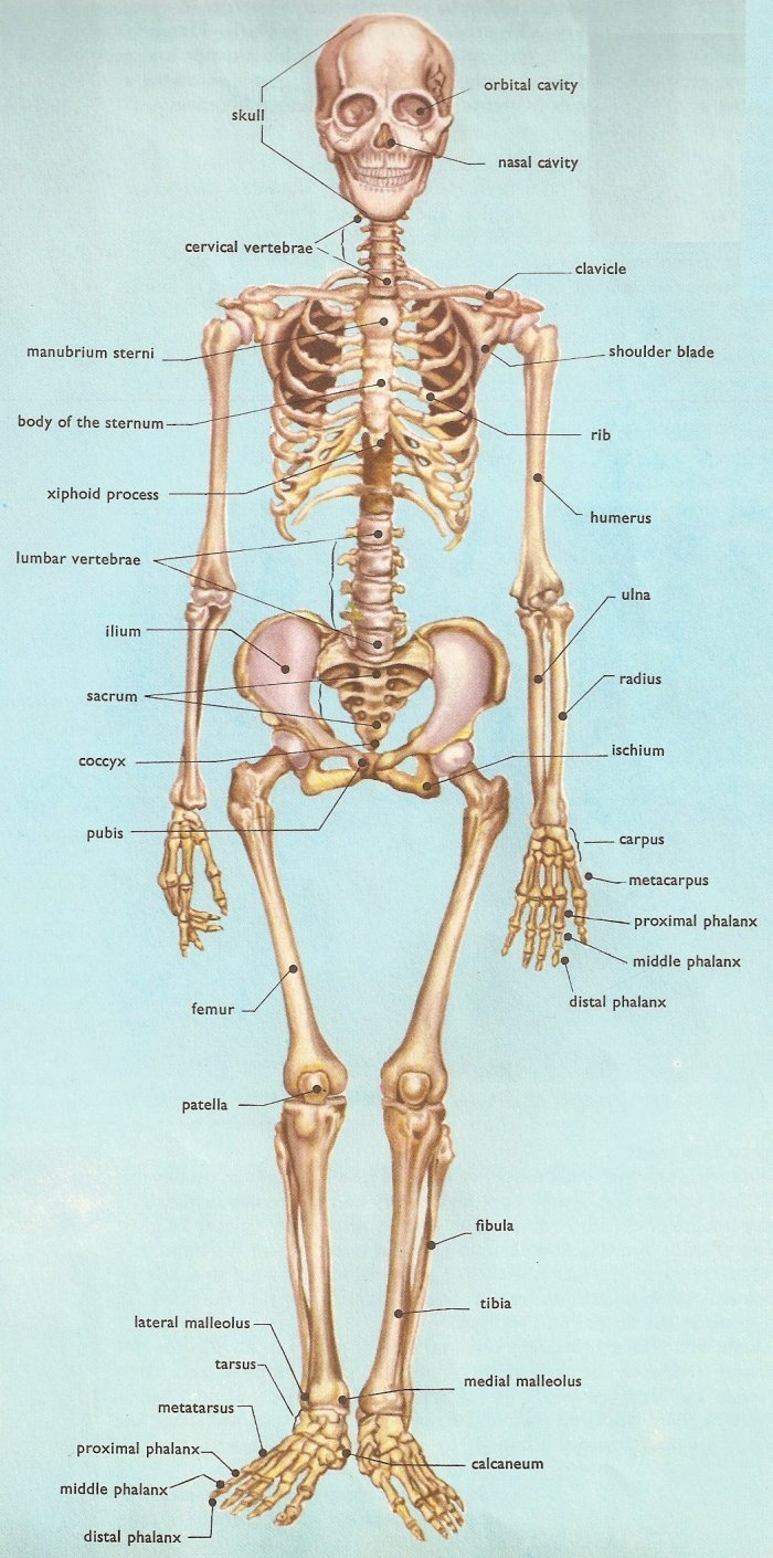
Images and X-rays
of Bone Metastases (normal
anatomy)
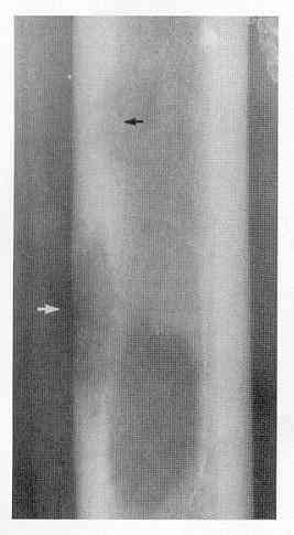
plain X-rays will show bone mets only if they are more advanced (go here)
-
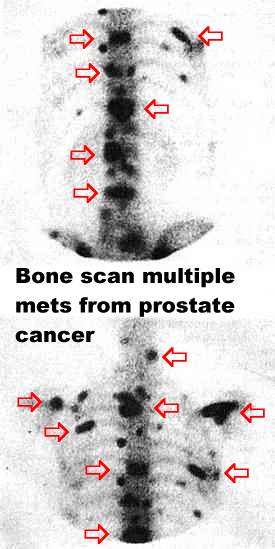
picture of a bone scan showing 'hot spots' (black areas) which are sites of bone cancer, see the close up of bone scan, and here and here
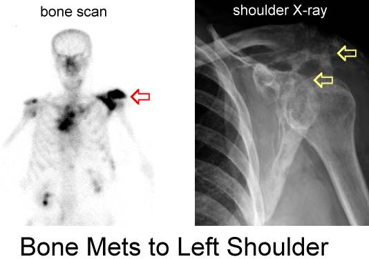
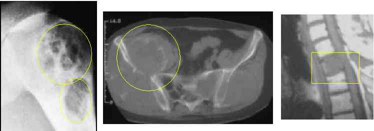
a bone scan or CT are more sensitive than plain Xrays (see right hip met and see rib mets).Advanced spine mets can lead to spinal cord syndrome go here.
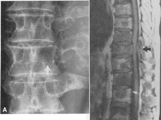
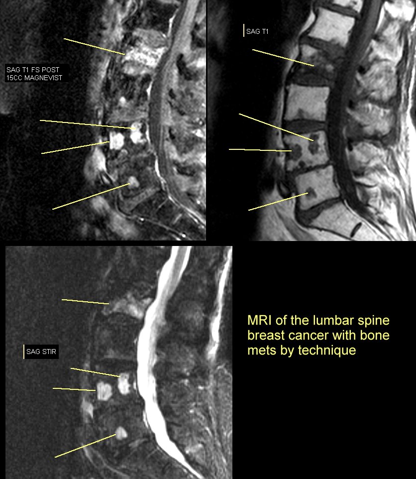
MRI is also more sensitive than plain Xrays. A woman had severe pain in her arm , the Xray was read as normal but the MRI was markedly abnormal (see here) she later developed a pathologic fracture see here. (other cases: see spine , spine, here and MRI) see MRI showing a pathologic fracture from lymphoma here. The specific MRI rechnique will influence the way bone mets appear (go here)
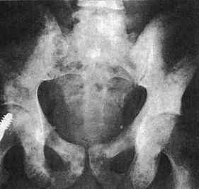
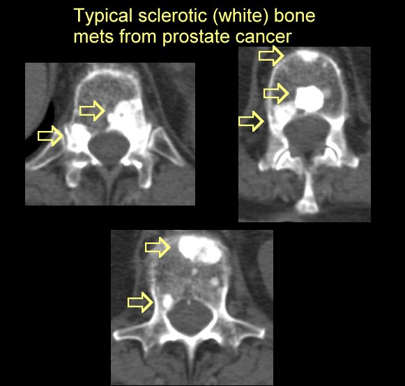
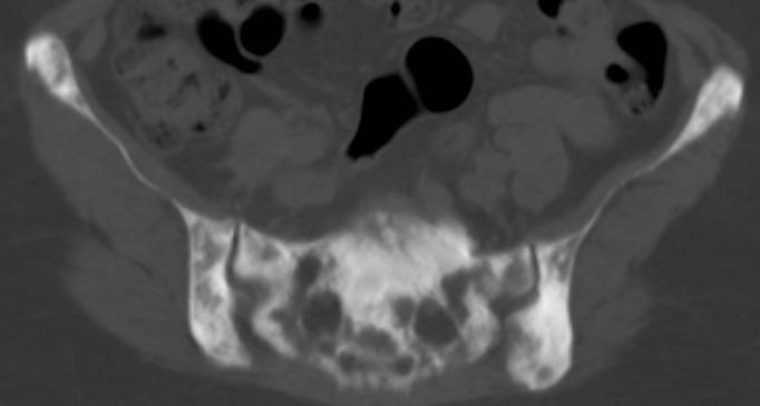
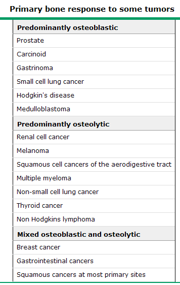
Some bone cancers make the bone look " too white" called osteoblastic mets of the pelvis or osteoblastic spine , CT of blastic mets here and here and here
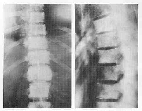
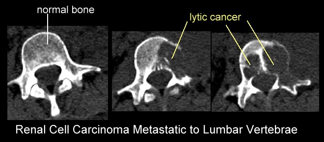
some cancer make "black holes" or lytic mets (another lytic lesion), renal cell cancer causes lytic lesions (go here), and see CT showing diffusely mottled bone here and here
more Xray/CT scan images,
PET scans are very good at showing bone mets go here
|
|
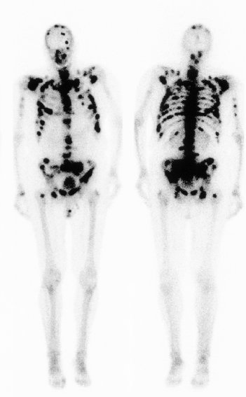
nuclear medicine bone scan showing multiple bones metastases (black areas) |
 |
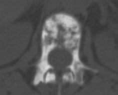
It is easy to over -
read an MRI and assume that any abnormal finding is serious. It's worth noting that most older adults have
abnormal MRI scans of their back that are entirely benign. Images of the Spine from Normal Volunteers. (see below) Herniation of the lumbar disk, as shown in Panel A, is found in 25 to 50 percent of asymptomatic subjects; extrusion of the disk material is found in 1 to 18 percent. Degeneration of the lumbar disk, shown in Panel B, increases with age and is found in 25 to 70 percent of asymptomatic subjects. Signal changes in the vertebral end plates (Panel C, arrows) are found in 10 percent of asymptomatic subjects; severe changes are found less frequently. Panel D shows a disk with a bright signal in the annular fissure. This represents degenerative changes that are found in 14 to 33 percent of asymptomatic subjects. Despite the high prevalence in healthy persons, these findings are often described as causing serious low back pain and are treated with spinal fusion.
|