| During the past two decades,
stereotactic radiosurgery has been widely used to treat
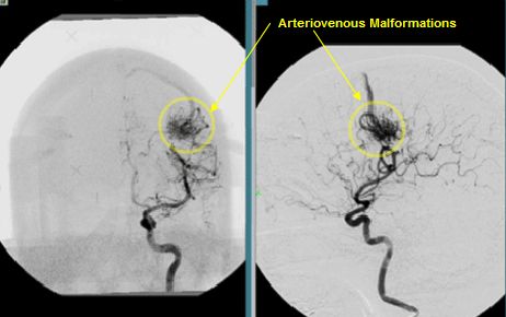
cerebral arteriovenous malformations,providing angiographic evidence of
cure (obliteration of the
malformation) in 80 to 95 percent of patients after a latency
period of three to five years. Hemorrhage has been
reported to occur in 2 to 5 percent of patients per year between
the time of radiosurgery and angiographic obliteration of the
malformation; however, it has been unclear whether — and
to what extent — the risk is reduced during this period as
compared with the risk before radiosurgery. The extent to which
the risk of hemorrhage is further reduced after angiographic
obliteration is also unclear. To address these questions, we
performed a retrospective study involving 500 patients who were
treated with stereotactic radiosurgery at our institute.
Background Angiography shows that stereotactic
radiosurgery obliterates most cerebral arteriovenous
malformations after a latency period of a few years. However,
the effect of this procedure on the risk of hemorrhage is
poorly understood.
Methods We performed a retrospective
observational study of 500 patients with malformations who were
treated with radiosurgery with use of a gamma knife. The rates
of hemorrhage were assessed during three periods: before
radiosurgery, between radiosurgery and the angiographic
documentation of obliteration of the malformation (latency
period), and after angiographic obliteration.
Results Forty-two hemorrhages were
documented before radiosurgery (median follow-up, 0.4 year), 23
during the latency period (median follow-up, 2.0 years), and 6
after obliteration (median follow-up, 5.4 years).
As compared with the period
between diagnosis and radiosurgery, the risk of hemorrhage
decreased by 54 percent during the latency period and by 88
percent after obliteration. The risk was significantly reduced
during the period after obliteration, as compared with the
latency period (hazard ratio, 0.26; 95 percent confidence
interval, 0.10 to 0.68; P=0.006). The reduction was greater
among patients who presented with hemorrhage than among those
without hemorrhage at presentation and similar in analyses that
took into account the delay in confirming obliteration by means
of angiography and analyses that excluded data obtained during
the first year after diagnosis.
Conclusions Radiosurgery significantly
decreases the risk of hemorrhage in patients with cerebral
arteriovenous malformations, even before there is angiographic
evidence of obliteration. The risk of hemorrhage is further
reduced, although not eliminated, after obliteration.
Discussion
We found that the risk of hemorrhage from
cerebral arteriovenous malformations was significantly
decreased after radiosurgery, both during the latency period
(between radiosurgery and angiographic obliteration) and after
angiographic obliteration. Previous studies have reported that
the risk of hemorrhage during the latency period decreases,
remains unchanged, or even increases, as compared with the
natural course of the disease. These studies tended to compare
the risk of hemorrhage among selected patients who underwent
radiosurgery with patients who did not undergo radiosurgery,
whereas we analyzed changes in the rate of hemorrhage relative
to the timing of radiosurgery in a large cohort of consecutive
patients.
Most previous studies assumed that
angiographic obliteration was the ultimate goal of
radiosurgery, because hemorrhage was rare once obliteration was
confirmed. Although recanalization of malformations can lead to
hemorrhages after obliteration, this phenomenon was not
observed in the six patients who had hemorrhage after
obliteration in our study. We found that a small risk of
hemorrhage remained after obliteration, although it was
markedly lower than that before radiosurgery.
We did not address the
mechanisms by which the risk of
hemorrhage may be reduced. However, histopathological
studies of arteriovenous malformations after radiosurgery
suggest potential mechanisms. Progressive thickening of the
intimal layer,which begins as early as three months after
radiosurgery, appears to decrease the stress to the vessel
walls. In addition, partial or complete thrombosis of the
irradiated vessels may decrease the number of patent vessels in
the malformation. In vessels with a decreased diameter,
thickening of the endothelium may cause occlusion at a
relatively early stage. When blood flow declines below the
threshold of detection by angiography, malformations, in
effect, become invisible (angiographic obliteration), although
they may still be evident histologically.
Our study has some limitations. Because we
did not include a control group of patients who did not undergo
radiosurgery, one concern is whether the decrease in the risk
of hemorrhage after radiosurgery reflects, at least in part,
the natural history of malformations, rather than effects of
the procedure itself. A natural decline in the rate of
recurrent bleeding has been reported within one year after the
rupture of arteriovenous malformations.
Because the criteria for conservative management were
not well described in previous reports of the natural history
of ruptured malformations,
it has remained unclear whether small malformations
that can be effectively treated with radiosurgery have a
similar natural decline in the rate of repeated hemorrhage.
However, the Cox models we used accounted for the time since
diagnosis. In addition, hemorrhage rates before radiosurgery in
our cohort appeared stable over a period of more than three
years after diagnosis, although the number of patients observed
for longer periods before radiosurgery was limited. In
addition, our results did not materially change in an analysis
that excluded data obtained during the first year after
diagnosis.
Another potential problem is the delay in
confirming angiographic obliteration.
The exact time of obliteration was not known but,
instead, was inferred on the basis of findings on consecutive
imaging studies. Angiography was initially carried out at six-month
intervals; after 1993, less invasive imaging was performed every
six months.
However, our results were materially unchanged
after adjustment for a potential delay of six months in identifying
obliteration. Although some patients had prior treatments, these
treatments are not expected to have a delayed effect, and the
results were more conservative when the period before these
treatments was excluded. Because our clinical practice incorporates
close follow-up of our patients according to standard schedules,
the retrospective nature of our analysis should not pose a problem.
The lack of blinding among those reviewing studies and judging
outcomes is also acceptable, since obliteration and hemorrhage
were diagnosed separately. There was some loss to follow-up,
but the assignment of extreme outcomes to these patients also
did not substantively affect the results of the analyses.
The gold standard for evaluating the effect
of radiosurgery on the risk of hemorrhage would be a randomized
trial comparing a group undergoing radiosurgery with a group
receiving no treatment. However, this approach is not possible,
because the beneficial effects of radiosurgery in terms of
angiographic cure are well recognized and hemorrhage is rare
after complete obliteration.
The large size and close follow-up of our cohort made it well
suited to an assessment of the outcomes of radiosurgery.
In conclusion, we found that the risk of
hemorrhage from cerebral arteriovenous malformations was
significantly reduced after stereotactic radiosurgery during
the latency period (after radiosurgery and before angiographic
obliteration) and that it was reduced even further after
obliteration. However, a risk of hemorrhage remained even after
malformations were no longer visible on imaging studies. |