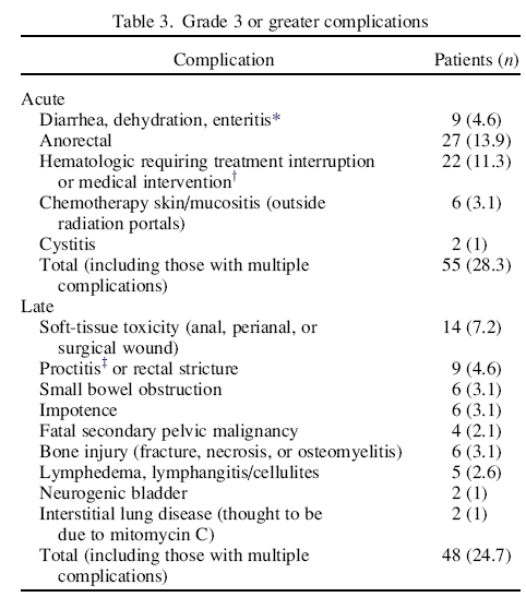
Grade 3 or greater late morbidities (according to the National Cancer Institute Common Toxicity Criteria; are summarized in Four patients died of a second malignancy in the pelvis (discussed in the last paragraph of this section); these were therefore considered possible treatment-related late fatalities. These were the only Grade 5 late morbidities. The most commonly encountered late morbidity was anorectal or perianal and included pain and/or soft-tissue breakdown and/or stricture. Of the 194 patients, 23 (12%) experienced anorectal late morbidity. This was significantly associated with the radiation dose to the anorectum. Of the 24 patients who received a boost to ≥55 Gy, 7 developed Grade 3 or greater anorectal morbidity (29%) compared with 16 (9%) of 170 treated to <55 Gy (p = 0.01, Fisher's exact test).
Two other late morbidities also appear to be associated with radiotherapy technique. Late small bowel obstruction (SBO) occurred in 6 patients. In all 6, the small bowel had been excluded after the first 30 Gy. Late bone injury (e.g., fracture, osteomyelitis, osteonecrosis) occurred in a different 6 patients.
With regard to SBO, excluding those patients who had undergone planned surgery (1 of whom had a SBO) and patients who were treated in the prechemotherapy era (none of whom had SBO), the SBO in the remaining patients appeared to be significantly sorted by radiotherapy technique: 4 (9%) of 44 patients treated with the anteroposterior–posteroanterior supine technique vs. 1 (0.7%) of 134 treated prone with multiple fields (p = .014, Fisher's exact test).
Bone injury was significantly associated with the radiation dose to the affected femoral head. The treatment policies and techniques that we used during the study period meant that patients with Stage N2, N3, or T4 cancer received a dose ≥44 Gy to the affected femoral head. The incidence of late bone injury was 9% (5 of 56) for those with a femoral head dose of ≥44 Gy vs. 1 (0.7%) of the 138 remaining patients (p = .008, Fisher's exact test).
Of the 194 patients, 56 (29%) had 68 additional malignancies. Of these patients, 36 had 44 additional malignancies that antedated their anal cancer, and 21 had 24 post-treatment additional malignancies (including 1 who also had had breast cancer before anal cancer). All but 7 of the post-treatment malignancies were outside the irradiated volume. Three cases of additional malignancy were considered to probably be radiation related: two in-field soft-tissue sarcomas (both cases treated in the prechemotherapy era) and one uterine sarcoma. One case of low-grade ureteral cancer probably was actually a synchronous malignancy, because the patient developed unilateral hydroureter requiring stenting shortly after completing treatment, with the diagnosis of ureteral malignancy not established until positive brushings were obtained 1 year later. The other three post-treatment pelvic malignancies were adenocarcinoma of the uterus, rectum, and prostate. Because the uterine adenocarcinoma was fatal, we scored it as one of the Grade 5 morbidities. However, this study had more uterine, rectal, and prostate adenocarcinomas occurring before anal cancer treatment than after (seven vs. three), suggesting that the 3 post-treatment cases were probably not radiation induced.
The analysis results of late morbidity in the present study have suggested methods to refine radiotherapy planning constraints. The substantial 29% late anorectal morbidity rate with a primary tumor dose of ≥55 Gy implies that dose heterogeneity within the boost planning target volumes (which, inevitably, will include the anus and low rectum) must be well controlled. The association of late SBO with anteroposterior–posteroanterior supine portals has led us to routinely use the same bowel exclusion techniques that we use for rectal cancer, a policy shift that has significantly reduced that morbidity. However, sparing of the femoral heads is difficult with conventional treatment planning techniques, particularly if the patient is treated prone (to help displace small bowel). The results of this study have demonstrated that patients with a femoral head dose of ≥44 Gy had a significantly increased incidence of late bone injury. These findings have led us to adopt intensity-modulated radiotherapy for all cases of anal cancer. Except for patients enrolled in RTOG studies, we have opted for two intensity-modulated radiotherapy plans: an initial 30 Gy to the pelvis and groin, followed by a boost of 16–30 Gy, with the boost dose depending on the tumor stage according to the policies described in the “Methods and Materials” section. Treatment planning constraints included requiring that the composite plan minimize the volume of small bowel receiving 30 Gy and the volume of femoral head receiving 44 Gy, while keeping the dose heterogeneity to <10% within the high-dose planning target volume.
|
|