Treatment
modalities
Combined surgery and radiation
Extrafascial hysterectomy after radiation therapy has been advocated for large IB
cervical cancer. Most reports have not identified a survival benefit attributable to the
addition of the hysterectomy to radiation therapy. The Gynecology Oncology Group (GOG
protocol #71) studied this question in a randomized trial. The adjuvant hysterectomy group
experienced a reduced rate of pelvic relapse compared to that seen in the group of
patients receiving radiotherapy alone. There was, however, no difference in overall
survival between the two groups Because of the increased complications associated
with extrafascial hysterectomy and the lack of evidence of any survival benefit, the ABS
does not recommend planned radiation therapy and adjuvant hysterectomy.
The use of adjuvant pelvic irradiation after radical hysterectomy and lymph node
dissection has been a controversial topic for decades. Most reports have not shown a
survival benefit in patients with risk factors for recurrence. Prognostic factors after
radical hysterectomy include lymph node status, tumor size, depth of invasion, and LVSI,
with positive pelvic lymph nodes being the strongest predictor of outcome.
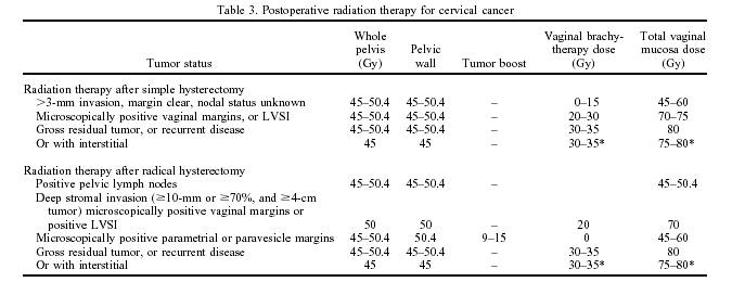
The ABS recommends adding postoperative radiotherapy for that
subgroup of patients that has at least two of the following risk factors for recurrence: (1)
greater than one-third stromal invasion, (2) LVSI, and (3) large (>4 cm)
tumor diameter. GOG #92 prospectively evaluated node-negative Stage IB cervical cancer
treated with radical hysterectomy and pelvic lymphadenectomy (±) pelvic radiotherapy in
this subgroup of patients. Those with extracervical spread or positive margins were
excluded. There was a statistically significant reduction in the overall recurrence rate
in the radiotherapy arm (15% vs. 28%) and an improvement in the recurrence-free rate at 2
years. Adjuvant pelvic radiotherapy after radical hysterectomy reduced the number of
recurrences (44% reduction). There was, however, a 6% incidence of Grades 3 and 4 toxicity
in that treatment arm compared to 2% in the surgery-only group.
The ABS recommends adding postoperative chemoradiotherapy for the
subgroup of patients with early-stage cervical cancer that meets at least one of the
following criteria: (1) positive pelvic lymph nodes, (2) positive
surgical margins, and (3) microscopic involvement of the parametrium. GOG #109
prospectively evaluated concurrent chemotherapy and pelvic radiotherapy or pelvic
radiotherapy alone after radical hysterectomy and pelvic lymphadenectomy in this subgroup.
Patients in the chemotherapy arm received bolus cisplatin and 96-h continuous infusion of
5-fluorouracil twice during irradiation and two additional courses after radiotherapy was
completed. There was a statistically significant improvement in the projected 4-year
progression-free interval (63% vs. 80%) and overall survival (71% vs. 81%) in favor of the
radiotherapy plus chemotherapy arm.The relative risk of death in this group was reduced by
50%.
Concurrent chemoradiation
The ABS recommends the addition of cis-platinum-based chemotherapy during the
course of definitive radiotherapy for patients with IB2–IVA disease. Definitive
irradiation has been the standard treatment for patients with advanced-stage disease
(IIB–IVA), as well as an excellent option for bulky early-stage cervical cancer. Five
randomized trials have recently demonstrated significant improvement in local control and
survival when concurrent chemotherapy was added to radiation therapy in patients with
early-stage disease and high risk for recurrence, as well as in patients with
advanced-stage disease.Four of these studies evaluated concurrent chemotherapy combined
with definitive radiotherapy.Radiation therapy recommendations
General
recommendations
The Patterns of Care studies have shown that recurrences and complications are
decreased when brachytherapy is used in addition to external beam irradiation (EBRT).
Therefore, the ABS strongly recommends that definitive irradiation for cervical carcinoma
include brachytherapy as a component. he ABS recommends limiting the total treatment
duration to less than 8 weeks when possible. Extending total treatment duration beyond 8
weeks can reduce local control and survival by about 1% per day of prolongation
Anemic patients should be transfused or receive erythropoietin to maintain the hematocrit
level above 30.
External beam therapy
The ABS recommends whole pelvic irradiation with four-field isocentric technique
with customized blocking, with all fields treated daily. The ABS recognizes that the
pelvic EBRT dose differs from institution to institution and can depend on the stage of
disease. Some institutions prefer to limit the whole pelvis dose for patients with early
disease and to perform the first intracavitary insertion after 20 Gy, with further EBRT
delivered with a central block in place. However, most institutions deliver 40–50 Gy
of EBRT to the entire pelvis. The inferior border should be determined clinically by
examining the vaginal extent of tumor. The anterior margin of the lateral portal
should include the pubis, so as to include the external iliac nodes, and the posterior
margin should extend to the sacral hollow to cover the uterosacral ligaments and internal
iliac nodes Some institutions use a midline block for part of the pelvic field irradiation
to shield the bladder and rectum to allow a higher dose to be given by brachytherapy.
There is no consensus regarding the use of midline blocks. The ABS recommends that, if
used, simple rectangular blocks should be 4 cm wide at midplane when intracavitary
brachytherapy applicators are used (Consensus Level 2). If EBRT doses greater than
45–50 Gy are to be given, the fields should be coned down after the initial
45–50 Gy to exclude small bowel. An additional parametrial boost may be delivered
with reduced portals to bring the EBRT contribution to the pelvic sidewall to
approximately 60 Gy when there is persistent parametrial tumor after whole pelvic EBRT.
Small bowel should be excluded from this boost volume as much as possible. If para-aortic
node metastases are present, the ABS recommends that the patient be treated with 45 Gy to
the para-aortic area, plus a 10–15-Gy boost to enlarged lymph nodes through reduced
lateral or rotational portals, along with chemotherapy (Consensus Level 2).
The ABS recommends use of two LDR insertions,
especially in larger tumors, to allow progressive tumor volume reduction and more
effective disease coverage with the second application. The first intracavitary insertion
is usually given after delivering 2 to 4 weeks of external beam irradiation as soon as
adequate pelvic geometry allows. The second application is usually performed 1 or 2 weeks
later such that the entire treatment course is completed within 8 weeks. Every attempt
should be made in the second application to replicate the position of the applicators in
the first implant, if the geometry was optimal. The ABS recognizes that, in certain
circumstances (unreliable patient, excellent geometry, small tumor volume), the
brachytherapy could be performed as a single insertion
- For modern applicators and using radiographic
localization, the ABS recommends the following alternative procedure
for locating point A, because of the considerations discussed above. Begin by
drawing a line connecting the middle of each of the colpostat sources (or the colpostat
capsules if the sources are not visible during localization) on the AP radiograph. Then,
from the intersection of this line with the tandem, move superiorly along the tandem 2 cm,
plus the radius of the colpostats, and then 2 cm perpendicular to the tandem in the
lateral directions. The dose shall be calculated and specified to point A on both the
right and left. The average of the right and left doses can be taken if a single point A
dose is needed.
For applications using a
tandem and vaginal cylinders, point A can be specified using the modified definition.
Begin at the flange on the tandem (indicating the external cervical os), travel superiorly
along the tandem 2 cm, then laterally perpendicular to the tandem 2 cm
- The ABS recommends reporting the dose at the
lateral vaginal surface (mucosa) (points Vs) and at 0.5 cm deep to the vaginal
surface (point Vd) and to correlate with clinical outcome.
- Nominal bladder point:
Practitioners should use the standard ICRU #38 definition for the bladder dose point, with
a minor change . The bladder point falls on the surface of a Foley balloon filled with 7
cc of iodinated radiographic contrast (diluted if necessary so as not to obscure the
localization markers on the AP radiograph) snugged into the trigone of the bladder. The
point selected should correspond to the maximum dose on the surface of this balloon. That
point may not be the most posterior aspect of the balloon, if it is situated to one side
or significantly superior or inferior to the vaginal applicator. It is to be noted that
the maximum bladder dose using three-dimensional dosimetry methods is usually higher than
the maximum bladder dose obtained by conventional methods
- Nominal rectum point:
The standard ICRU #38 definition of rectal point (0.5 cm posterior to the posterior
vaginal wall as identified by radiopaque gauze in the vagina) can be used, because it is
easy to determine and will maintain standardization with common practice. Additional
information regarding the anterior rectal wall may be obtained by injecting a diluted
solution of barium contrast into the rectum. Attention should also be given to
radiographic visualization and dose to the sigmoid colon, because it may pass close to the
tandem. Alternate localization tools, such as lead markers in a catheter or measurement
devices, are not recommended, because they often lie much posterior to the anterior wall
and therefore result in erroneous low point doses.
- The dose to the nominal rectal and bladder points
should be kept as low as possible, although consistent with delivering appropriate tumor
doses. Every effort should be made to keep the bladder dose to
<90% of point A dose, the total bladder dose below 80 Gy, and the total rectal dose
below 75 Gy.
|